Thomas Lilieholm1, Matt Henningsen2, Azam Ahmed3, Alan McMillan1,4, and Walter F Block1,4,5
1Medical Physics, University of Wisconsin at Madison, Madison, WI, United States, 2Electrical Engineering, University of Wisconsin at Madison, Madison, WI, United States, 3Neurological Surgery, University of Wisconsin at Madison, Madison, WI, United States, 4Radiology, University of Wisconsin at Madison, Madison, WI, United States, 5Biomedical Engineering, University of Wisconsin at Madison, Madison, WI, United States
1Medical Physics, University of Wisconsin at Madison, Madison, WI, United States, 2Electrical Engineering, University of Wisconsin at Madison, Madison, WI, United States, 3Neurological Surgery, University of Wisconsin at Madison, Madison, WI, United States, 4Radiology, University of Wisconsin at Madison, Madison, WI, United States, 5Biomedical Engineering, University of Wisconsin at Madison, Madison, WI, United States
ML-driven autonomous segmentation of intracerebral hemorrhages (ICH) can be used to quantify hematoma volume for use in image-guided minimally-invasive surgical evacuation. We built a model to detect and segment clot and edema from ICH MR scans.
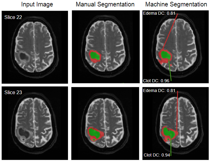
(Left) input T2-W images, (middle) manual segmentation, (right) automatic segmentation of a case from the testing dataset. Green and red regions correspond to tissue identified as clot and edema, respectively, and demonstrate good agreement between the manual and automated segmentations.
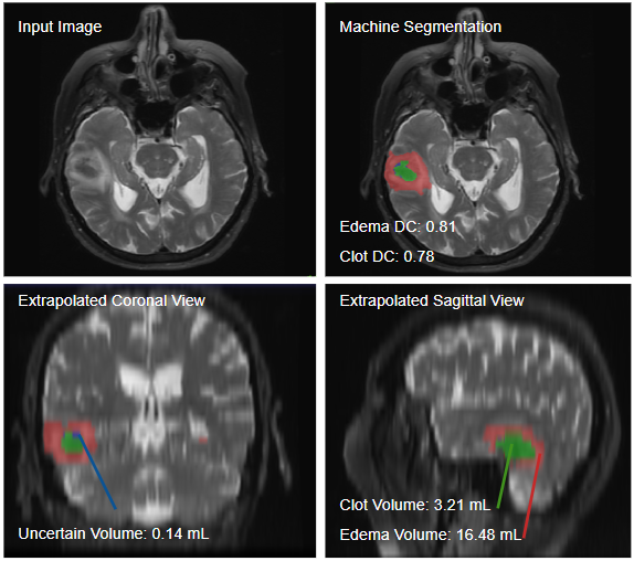
After autonomous-generation of a set of clot and edema segmentations in the axial view, coronal and sagittal views can be extrapolated from the known dimensions of the original dataset. This allows for better visualization and localization of clot components, and their relevant volumes. The small blue region is uncertain- the model identifies it as either clot or edema, but cannot distinguish between the two categories.