Nate Tran1,2, Tracy Luks1, Devika Nair1, Angela Jakary1, Yan Li1, Janine Lupo1, Javier Villanueva-Meyer1, Nicholas Butowski3, Jennifer Clarke3, and Susan Chang3
1Department of Radiology & Biomedical Imaging, University of California, San Francisco, SAN FRANCISCO, CA, United States, 2UCSF/UC Berkeley Graduate Program in Bioengineering, SAN FRANCISCO, CA, United States, 3Department of Neurological Surgery, University of California, San Francisco, SAN FRANCISCO, CA, United States
1Department of Radiology & Biomedical Imaging, University of California, San Francisco, SAN FRANCISCO, CA, United States, 2UCSF/UC Berkeley Graduate Program in Bioengineering, SAN FRANCISCO, CA, United States, 3Department of Neurological Surgery, University of California, San Francisco, SAN FRANCISCO, CA, United States
We
trained and tested random forest models using metabolic, perfusion, and
diffusion images at both preRT and midRT scans, and found that not confining
these metrics to the anatomical lesion boundaries improved outcome prediction.
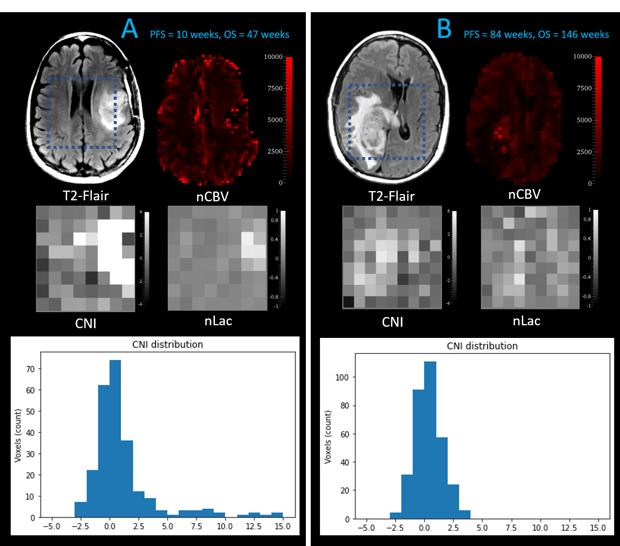
Figure 1: Patient B has a CEL volume of 28.4 cm3,
progressed at 84 weeks, and died at 146 weeks. Although Patient A has smaller
CEL & T2L volumes (CEL=10.9 cm3), they progressed much sooner at
10 weeks, and died at only 47 weeks
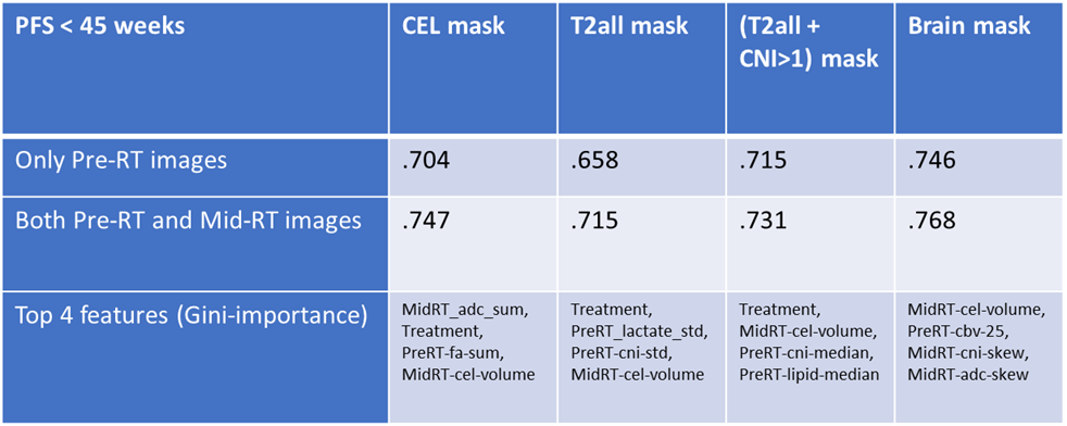
Table 1: Performance of RandomForest model to predict
whether or not OS<45 weeks for each mask using just pre-RT images, and both
pre-RT and mid-RT images