René Schranzer1,2, Steffen Bollmann3, Simon Hametner2, Christian Menard1, Siegfried Trattnig4, Fritz Leutmezer2, Paulus Stefan Rommer2, Thomas Berger2, Assunta Dal-Bianco2, and Günther Grabner1,2,4
1Department of Medical Engineering, Carinthia University of Applied Sciences, Klagenfurt, Austria, 2Department of Neurology, Medical University of Vienna, Vienna, Austria, 3School of Information Technology and Electrical Engineering, The University of Queensland, Brisbane, Australia, 4Department of Biomedical Imaging and Image-guided Therapy, High Field Magnetic Resonance Centre, Vienna, Austria
1Department of Medical Engineering, Carinthia University of Applied Sciences, Klagenfurt, Austria, 2Department of Neurology, Medical University of Vienna, Vienna, Austria, 3School of Information Technology and Electrical Engineering, The University of Queensland, Brisbane, Australia, 4Department of Biomedical Imaging and Image-guided Therapy, High Field Magnetic Resonance Centre, Vienna, Austria
We developed a pipeline, based on neural
networks, that provides high quality lesion segmentation and automatic
classification of MS lesions based on the presence or absence of an iron-rim.
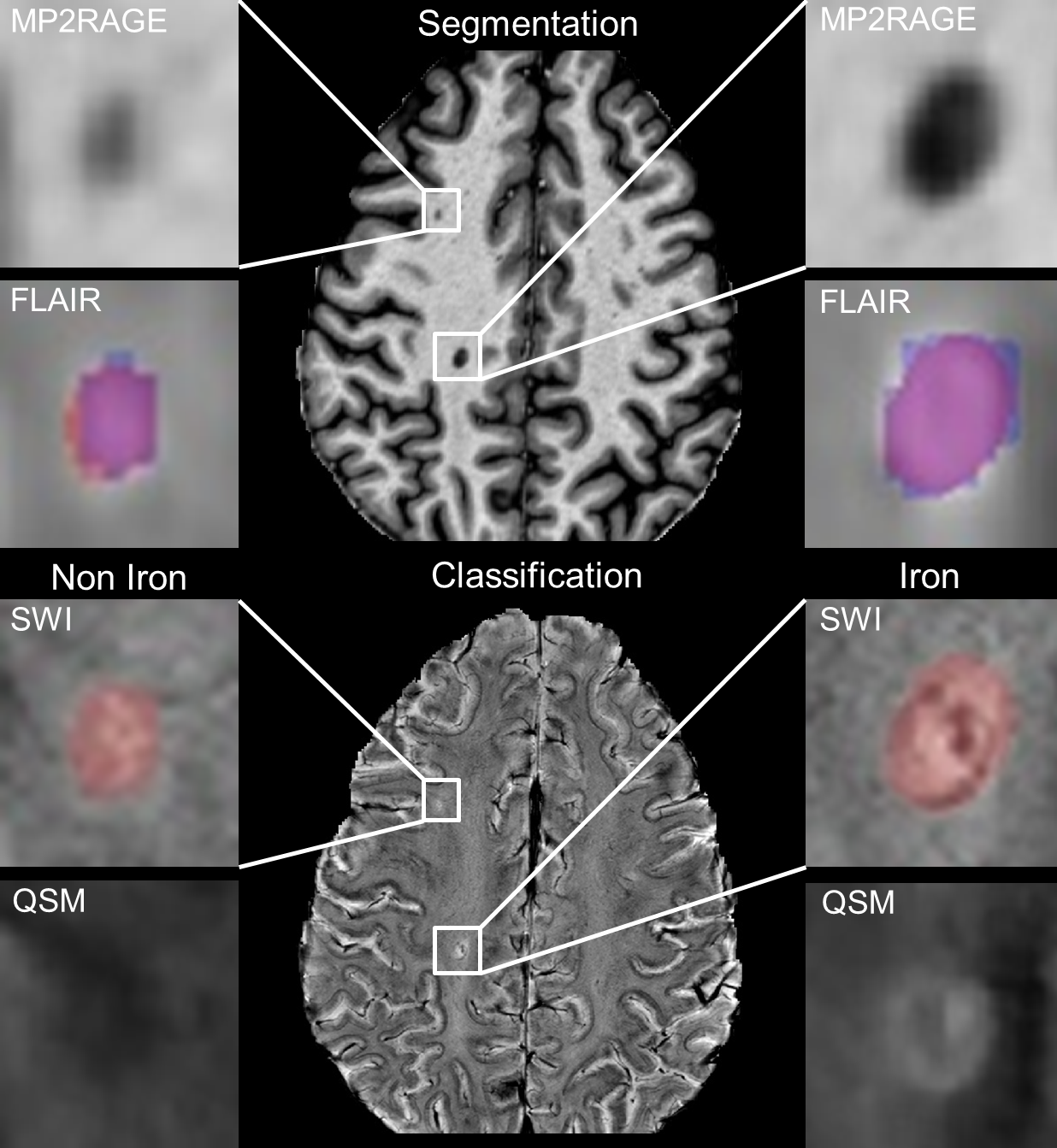
Figure
1.: Lesion segmentation and iron classification
results of the CNNs from the same slice of one representative MS patient: A
segmentation comparison for two MS lesions, between manual expert labeling
(blue) and CNN labeling (red) is shown in the top image. An example for a
non-iron (left) and iron lesion (right) classification from the same area as above is
shown on the bottom. A prominent hypointense and hyperintense iron-rim is
visible in the SWI and QSM image, respectively.
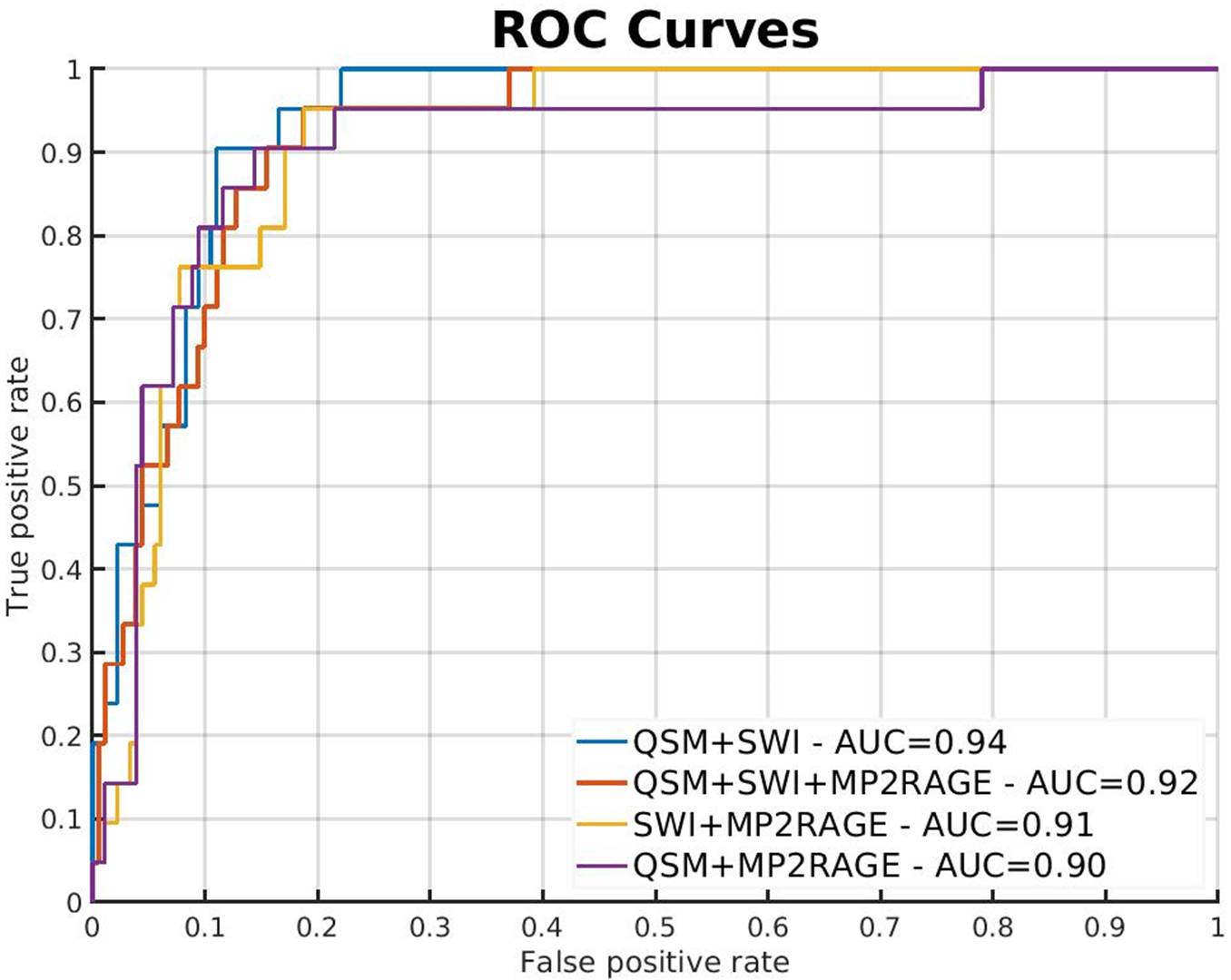
Figure 2.: Receiver operating characteristic curves for all network
combinations: The Graph shows ROC curves with true-positive rate plotted against
false-positive rate for lesion-wise prediction of iron.