Margaux Verdier1,2, Justine Belko1, Jeremy Deverdun1, Nicolas Menjot de Champfleur1,3, Thomas Troalen2, Bénédicte Maréchal4,5,6, Emmanuelle Le Bars1, and Till Huelnhagen4,5,6
1I2FH , Neuroradiology, CHU Montpellier, Montpellier University, France, Montpellier, France, 2Siemens Healthcare, Saint Denis, France, 3Laboratoire Charles Coulomb, University of Montpellier, France, Montpellier, France, 4Advanced Clinical Imaging Technology, Siemens Healthcare AG, Lausanne, Switzerland, 5LTS5, Ecole Polytechnique Fédérale de Lausanne, Lausanne, Switzerland, 6Radiology Department, Lausanne University Hospital and University of Lausanne, Switzerland, Lausanne, Switzerland
1I2FH , Neuroradiology, CHU Montpellier, Montpellier University, France, Montpellier, France, 2Siemens Healthcare, Saint Denis, France, 3Laboratoire Charles Coulomb, University of Montpellier, France, Montpellier, France, 4Advanced Clinical Imaging Technology, Siemens Healthcare AG, Lausanne, Switzerland, 5LTS5, Ecole Polytechnique Fédérale de Lausanne, Lausanne, Switzerland, 6Radiology Department, Lausanne University Hospital and University of Lausanne, Switzerland, Lausanne, Switzerland
Convolutional neural network correctly
segments low-grade gliomas using common clinical T1 and T2-FLAIR sequences,
facilitating efficient tumor growth evaluation. Segmentation errors can
occur when gliomas show strong heterogeneous intensity patterns.
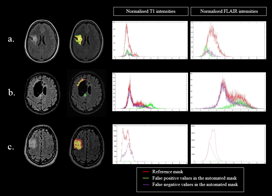
Figure 3: Three
different profiles (left column) with the native FLAIR images and the segmentation masks overlaid on FLAIR
images; reference mask in red, automated mask in green and common area in yellow.
Corresponding histograms of the normalized T1 and FLAIR signal intensities with
manual mask (red), false positive values in the automated mask (green), and false
negative values in the automated mask (purple). a : Best automated segmentation;
b : Poorest automated segmentation; c : Moderate automated segmentation.
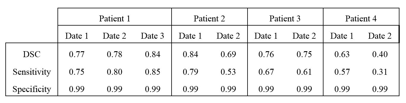
Table 2: Performance of the automated tumor segmentation in
the test patients.