Ozan Genc1, Sivakami Avadiappan1, Yicheng Chen2, Christopher Hess1, and Janine M. Lupo1
1Department of Radiology and Biomedical Imaging, University of California San Francisco, San Francisco, CA, United States, 2Facebook Inc., Mountain View, CA, United States
1Department of Radiology and Biomedical Imaging, University of California San Francisco, San Francisco, CA, United States, 2Facebook Inc., Mountain View, CA, United States
Synthetic SWI data was generated from T2* magnitude images using an LSGAN deep learning model. Findings suggest that our deep learning model is able to improve microbleed contrast on T2* magnitude images.
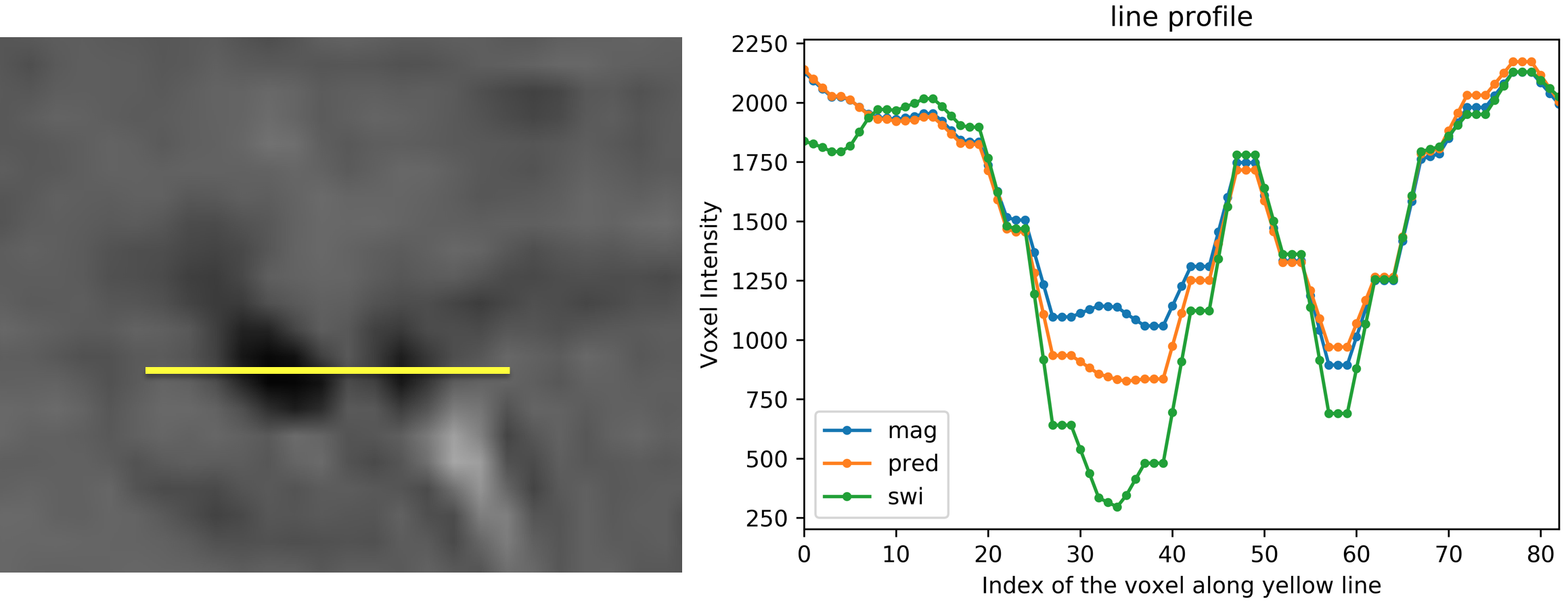
On the left, two CMBs are shown on a SWI image. On the right, voxel intensities of original SWI (green), predicted SWI (orange) and magnitude image (blue) along the yellow line are shown.
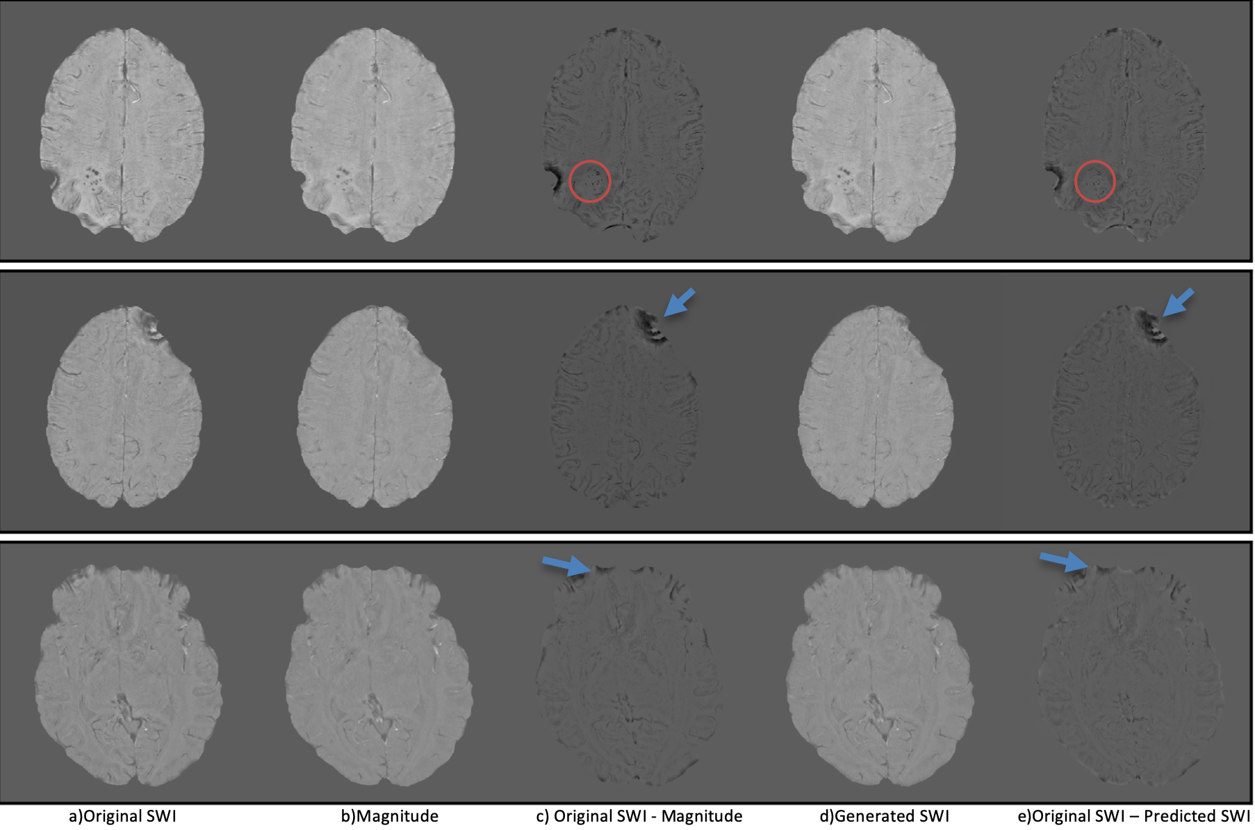
(a) Original SWI, (b) magnitude image, (c) difference image of original SWI and magnitude image, (d) predicted SWI, (e) difference image of original SWI and predicted SWI. Red circles show CMBs in difference images. Blue arrows show phase artifacts in difference images.