Bobby Runderkamp1, Thomas Roos2, Wietske van der Zwaag2, Matthan Caan3, Gustav Strijkers3, and Aart Nederveen1
1Radiology and Nuclear Medicine, Amsterdam UMC, Amsterdam, Netherlands, 2Spinoza Center for Neuroimaging, Amsterdam, Netherlands, 3Department of Biomedical Engineering and Physics, Amsterdam UMC, Amsterdam, Netherlands
1Radiology and Nuclear Medicine, Amsterdam UMC, Amsterdam, Netherlands, 2Spinoza Center for Neuroimaging, Amsterdam, Netherlands, 3Department of Biomedical Engineering and Physics, Amsterdam UMC, Amsterdam, Netherlands
We
used multi-channel parallel transmit to address B1+ inhomogeneity in the liver,
comparing phase shimming with a kt-points pulse. In volunteers with a small waist, kt-points and phase shimming
can homogenize B1+ in a considerable part of the liver.
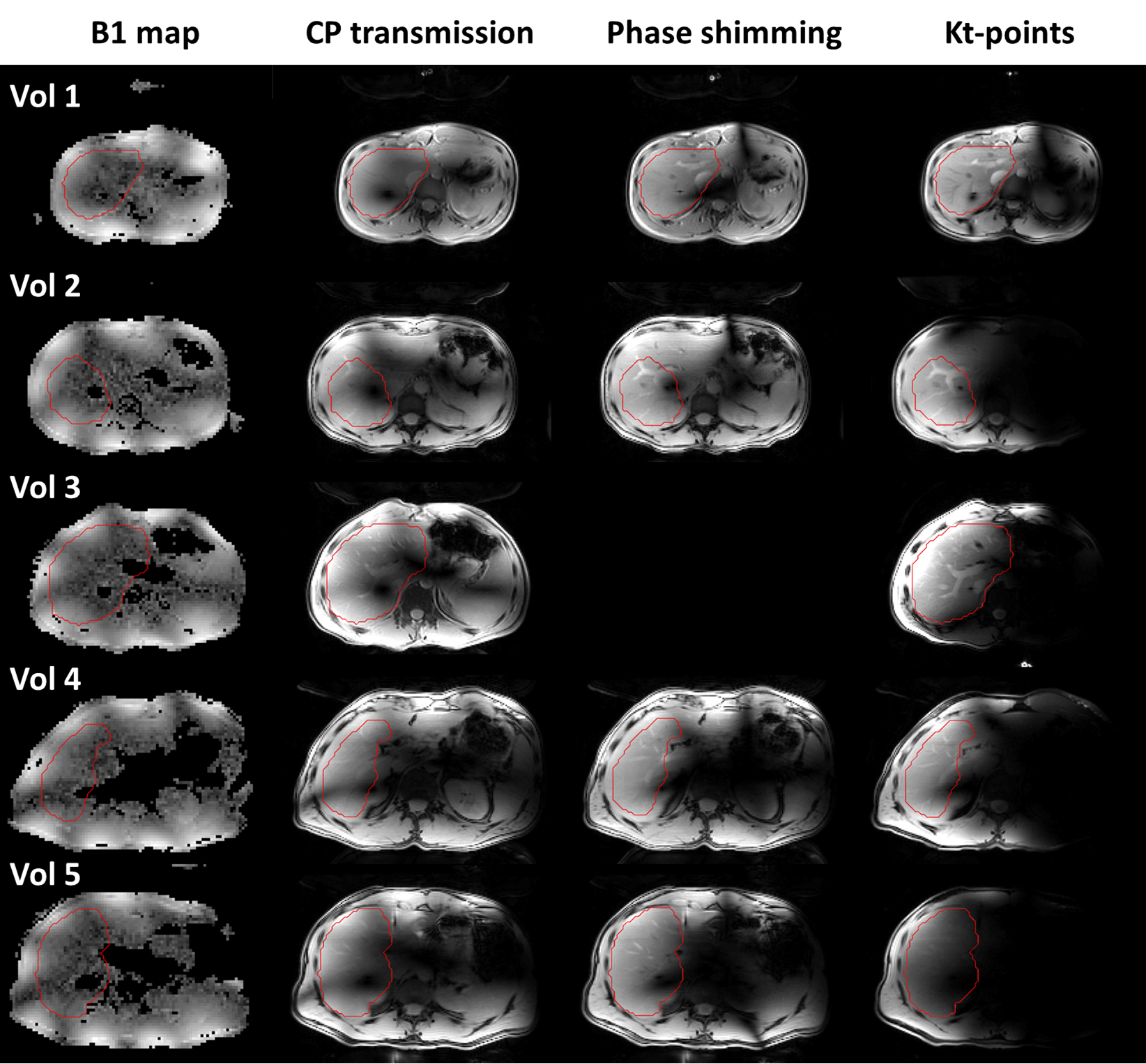
Figure 2: B1-maps
and magnitude images acquired with CP-transmission, phase shimming and kt-points,
at the level of the right portal vein, for all volunteers. The drawn shim VOI
in this slice is outlined in red. For volunteer 3, no phase shimmed image was
acquired. In the smallest volunteers, 1&2, phase shimming and kt-points
show considerable signal and homogeneity increase compared to CP-transmission. In the larger volunteers,
4&5, B1-maps appear more severely noise-clipped, resulting in lower to no
improvement using kt-points and phase shimming.
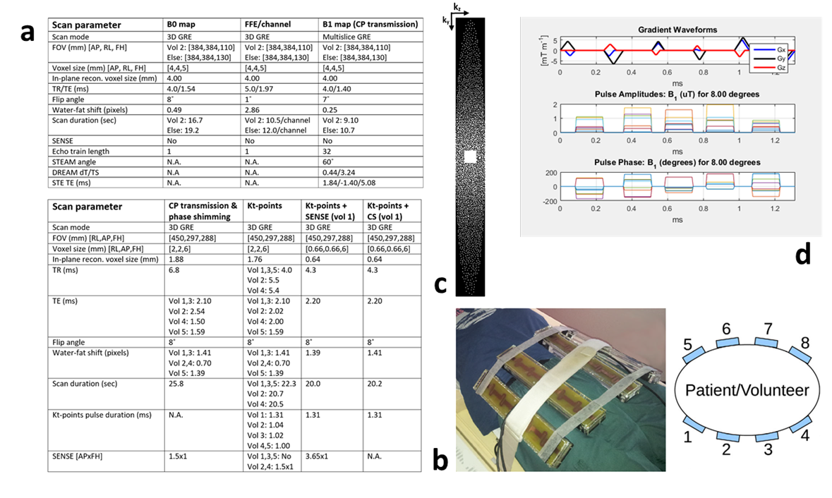
Figure 1: a)Parameters for the scans used for calculation of optimized phase offsets and
kt-points pulse, and the eventually B1+ homogenized scans. For kt-points scans, the echo
time was defined as the time between the echo and the middle of the kt-points pulse. b)The 8-channel fractionated dipole antenna body array used
for parallel transmission. In this study, the antennas were placed
asymmetrically towards the side of the body containing the liver. c)The
undersampling k-space pattern used in the CS scan. d)An exemplary kt-point pulse trajectory and pulse amplitudes and
phases.