Andreas Georg Berg1,2, Stefan Hengsbach3, and Klaus Bade3
1Center for Medical Physics and Biomedical Engineering High field MR-Center, Medical University of Vienna, Vienna, Austria, 2High-Field MR-Center (MRCE), Medical Universits of Vienna, Vienna, Austria, 3Karlsruhe Nano Micro Facility (KNMF), Karlsruhe Institute of Technology (KIT), Leopoldshafen-Eggenstein, Germany
1Center for Medical Physics and Biomedical Engineering High field MR-Center, Medical University of Vienna, Vienna, Austria, 2High-Field MR-Center (MRCE), Medical Universits of Vienna, Vienna, Austria, 3Karlsruhe Nano Micro Facility (KNMF), Karlsruhe Institute of Technology (KIT), Leopoldshafen-Eggenstein, Germany
Cube set for the measurement of spatial
resolution for 3 different orthogonal spatial encodings, for preclinical and special
High-Field human MR-scanners. The design features 3D-periodic structures (a=128µm
- 4µm): manufacturing technology, prototype phantom, exemplary MR evaluation.

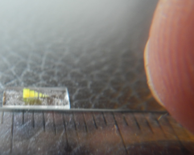
Fig. 1a Photographic image of the cube set on a carrier glass platelet.
The set of cubes comprises
periodic openings in the massive resist material in all of the 3 different
spatial directions. Each cube is characterized by a decreasingly less spatial
period for the evaluation of the spatial resolution ai = 128, 96, 64,
48, 32, 24, 16, 8, 4 µm (cf. fig. 1b right). Carrier layer: width/length [mm]: 2.8/7.

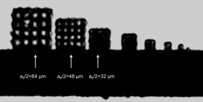
Fig. 2 High resolution sagittal image taken form the 3D-data set of a T2*w
gradient-echo sequence (VS: 31x31x30 µm3, Mtx: 128x128x36). The grid
structure of the cubes with spatial period a3/2 = 64 µm (leftmost) and
a4/2=48 µm (2nd from left) can be clearly resolved.
However the cavities in the cube a5/2 = 32 µm can hardly be observed any more.
This cube is not resolved indicating spatial resolution worse than the nominal
voxel size (≈30 µm). TR: 100 ms; TE: 7.8 ms; bw: 110 Hz/px.
