Christoph Leuze1, Supriya Sathyanarayana1, Bruce L Daniel1, and Jennifer A McNab1
1Radiology, Stanford University, Stanford, CA, United States
1Radiology, Stanford University, Stanford, CA, United States
We present a method for alignment of augmented reality display
of brain MRI with the patient’s real-world head with potential applications to
an AR-neuronavigation system that relies on a see-through display.
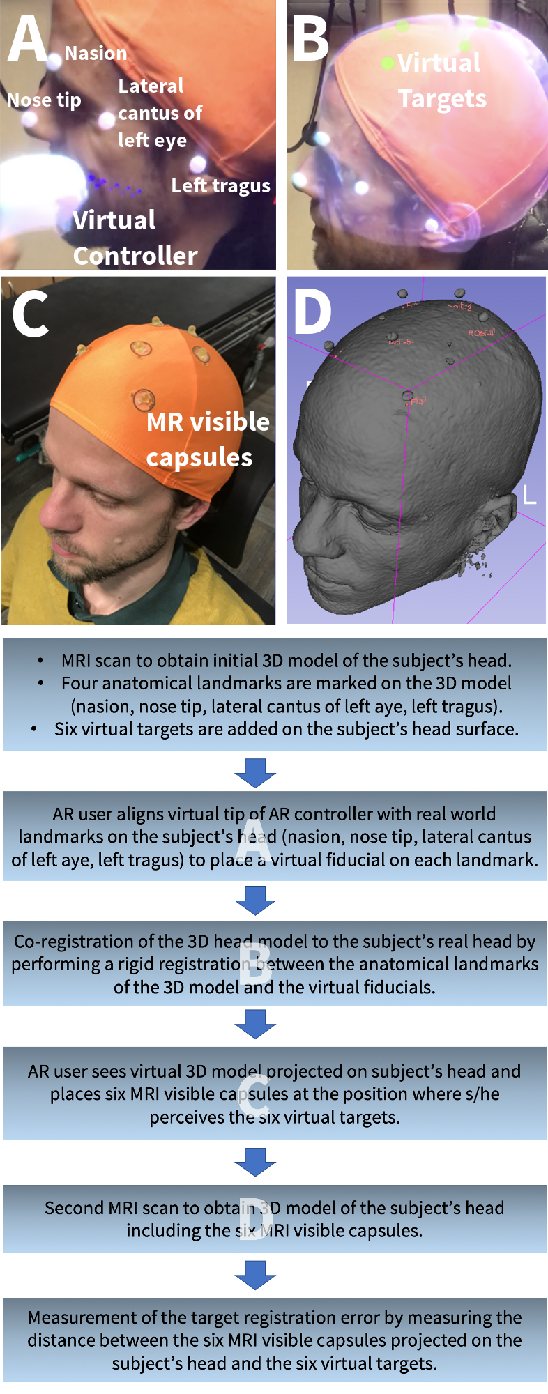
(Top) A View through the MagicLeap. Using
the virtual rendering of the MagicLeap controller, the AR user placed virtual
fiducials at anatomical landmarks around the subject’s head. B The MRI
head rendering overlaid on a subject’s head with the virtual targets in green. C
The AR user attached MRI visible capsules to the subject’s head at the
positions where s/he perceived the virtual targets. D A surface
rendering of the second MRI scan showing the capsules on the
subject’s head. (Bottom) A flowchart of the complete alignment and accuracy
measurement procedure (A-D refer to top image).
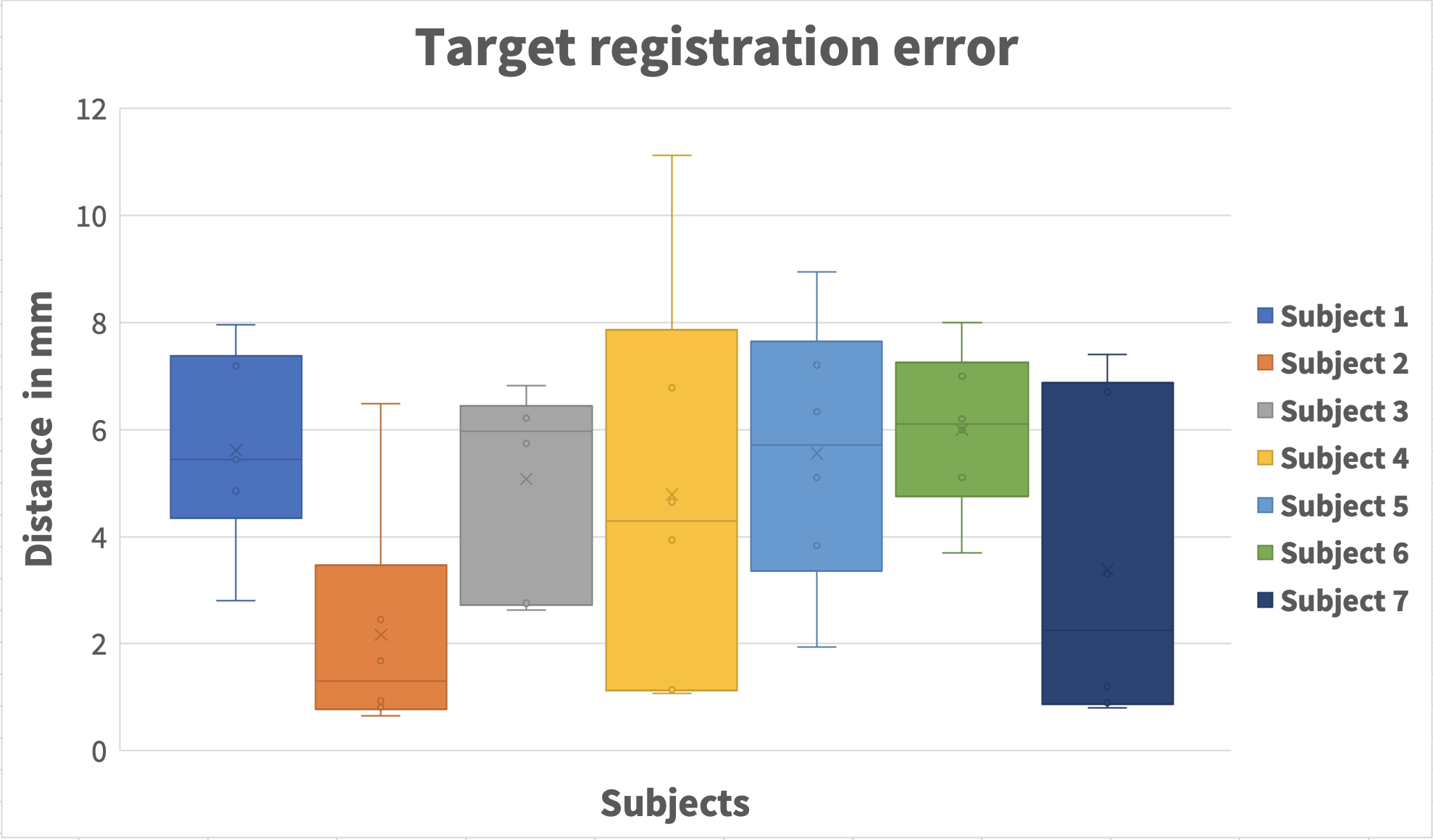
The TRE for all seven subjects measured as the distance
between the center of the MRI visible capsule projected to the head
surface and the original target projected on the head surface. We measured a
mean TRE = 4.7 ± 2.6 mm.