Philip Meng-en Lee1, Hsin-Yu Chen1, Jeremy W Gordon1, Zihan Zhu1, Peder EZ Larson1, Nicholas Dwork1, Mark Van Criekinge1, Lucas Carvajal1, Michael A Ohliger1, Zhen J Wang1, Duan Xu1, John Kurhanewicz1, Robert A Bok1, Rahul Aggarwal2, Pamela N Munster2, and Daniel B Vigneron1
1Department of Radiology & Biomedical Imaging, University of California, San Francisco, San Francisco, CA, United States, 2Department of Medicine, University of California, San Francisco, San Francisco, CA, United States
1Department of Radiology & Biomedical Imaging, University of California, San Francisco, San Francisco, CA, United States, 2Department of Medicine, University of California, San Francisco, San Francisco, CA, United States
A voxel-wise transmit B1 field correction method for surface coil hyperpolarized 13C MRSI scans was developed and implemented within a denoising pipeline, yielding more accurate quantification of pyruvate to lactate conversion rates, kPL, as validated by simulations.
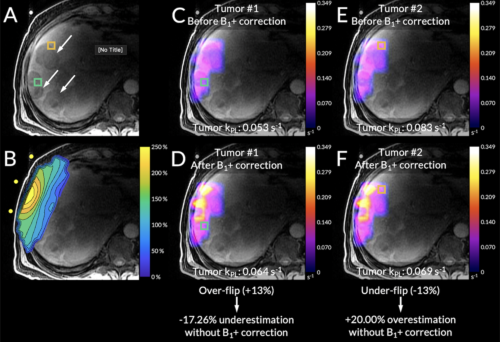
Figure 3. From Scan #3, (A) an axial T1-weighted spoiled gradient-echo anatomical scan. (B) The B1+ profile aligned to its position during the scan using the coil’s fiducial markers. (C-D) The kPL maps before and after B1+ correction with an over-flipped tumor voxel highlighted in green. (E-F) The kPL maps before and after B1+ correction with an under-flipped tumor voxel highlighted in orange.
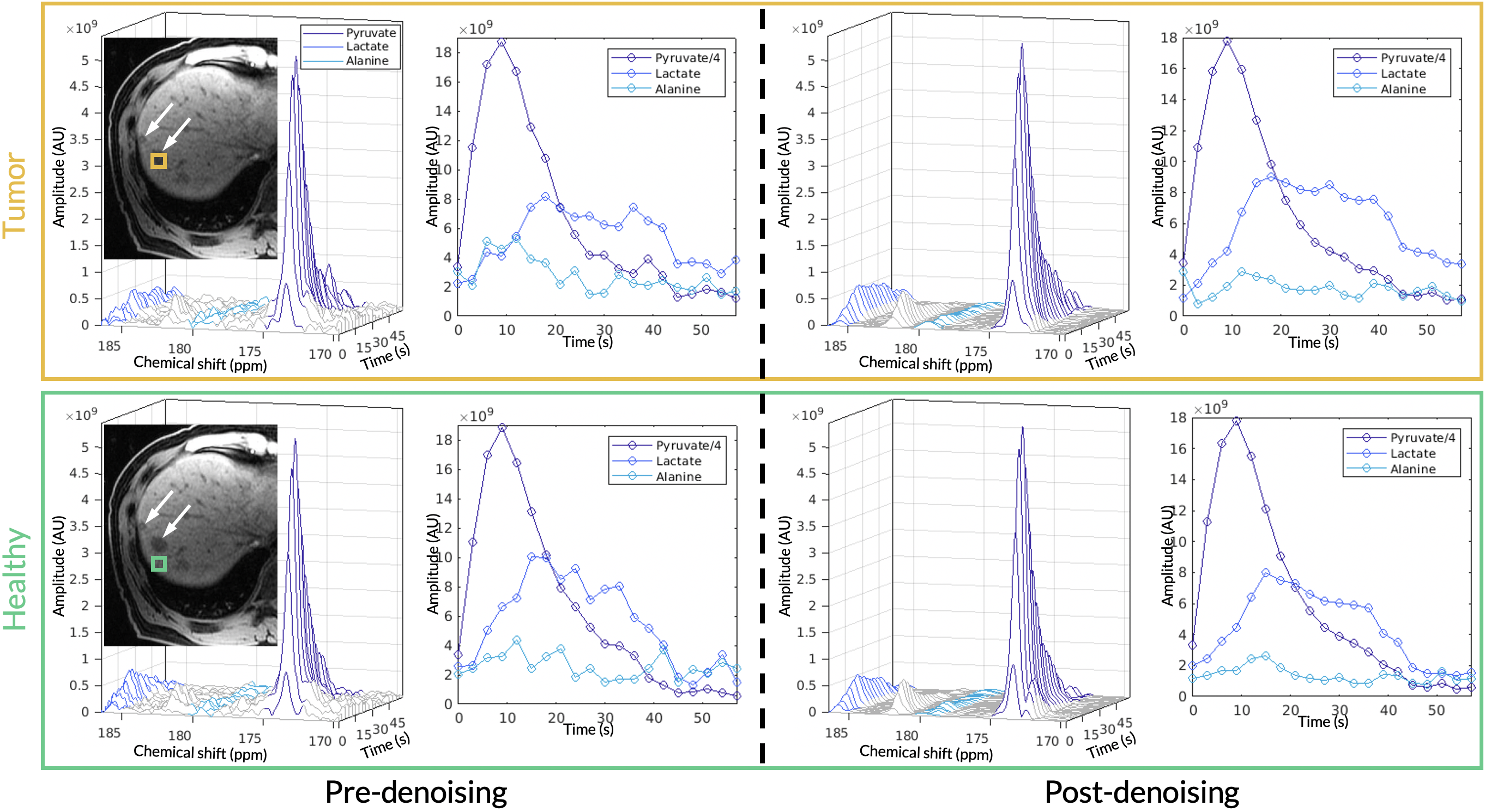
Figure 2. The top panel (orange box) shows the acquired HP 13C EPSI spectrum and dynamics before and after denoising for the tumor voxel. Likewise, the bottom panel (green box) shows the acquired EPSI spectrum and dynamics before and after denoising for a healthy-appearing voxel. The adjacent T1-weighted anatomical images highlight locations of the metastases and the selected voxel. Both are representative voxels taken from Scan #5.