Leo Konst Marecki1, Eric Konst Marecki1, and Xiaoliang Zhang1
1Biomedical Engineering, SUNY University at Buffalo, Buffalo, NY, United States
1Biomedical Engineering, SUNY University at Buffalo, Buffalo, NY, United States
Liquid
phantom material enclosed in 3D printed Nylon containers can be used to
generate the geometry and tissue characteristics of the human knee. This enables SAR modeling of each organ to
determine heat deposition in a 10 Tesla MRI.
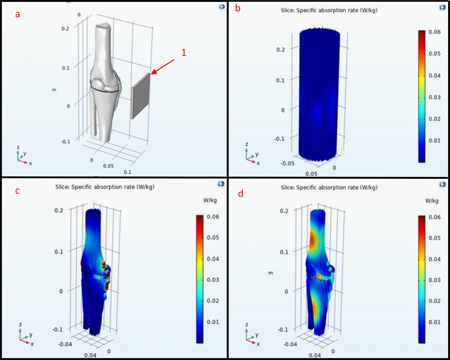
Figure 4:
Image a is the 3D knee model and orientation of each test to the antenna
(1). Image b is the water only based
phantom and shows a uniform SAR at all height, angle, and radius. Image c is the SAR of the knee phantom and
shows a large SAR on the MCL with a low SAR on every other component. Image d is the knee phantom placed into the
water cylinder in figure b and shows increased SAR in the cartilage and
anterior locations of the femur and tibia.
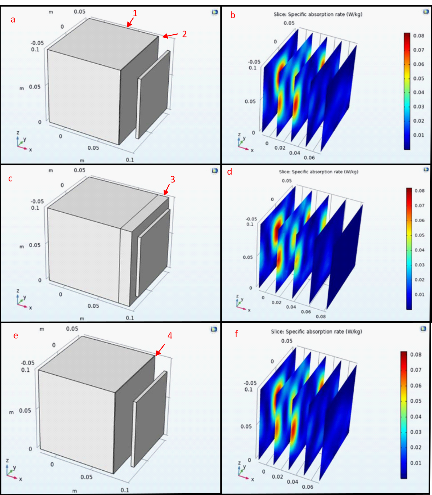
Figure 2: SAR
Profiles of 0.1 meter edge length water solutions (1) being excited by an
antenna at 425 MHz(2). In images c-d a 8
mm nylon rectangle is between the water and the antenna (3) and a 0.8 mm
rectangle(4) in figures e-f. When
compared to image b the SAR maps d and f show that when Nylon is 8 cm or 0.8 mm
the Nylon decreases the Maximum SAR in the water in no noticeable way. The SAR in the image d model shows that the
SAR in the Nylon is very low and will prevent heating of the Nylon.