Rita Schmidt1,2, Ghil Jona3, and Edna Furman-Haran2,3
1Neurobiology, Weizmann Institute of Science, Rehovot, Israel, 2The Azrieli National Institute for Human Brain Imaging and Research, Weizmann Institute of Science, Rehovot, Israel, 3Life Sciences Core Facilities, Weizmann Institute of Science, Rehovot, Israel
1Neurobiology, Weizmann Institute of Science, Rehovot, Israel, 2The Azrieli National Institute for Human Brain Imaging and Research, Weizmann Institute of Science, Rehovot, Israel, 3Life Sciences Core Facilities, Weizmann Institute of Science, Rehovot, Israel
In this study, a 3D-printed head-shaped phantom
with brain mimicking metabolites and lipid layer examined for 7T MRI and MRSI. The
phantom was designed to resemble the brain with respect to B0 and B1
distributions, metabolites and lipid layer.
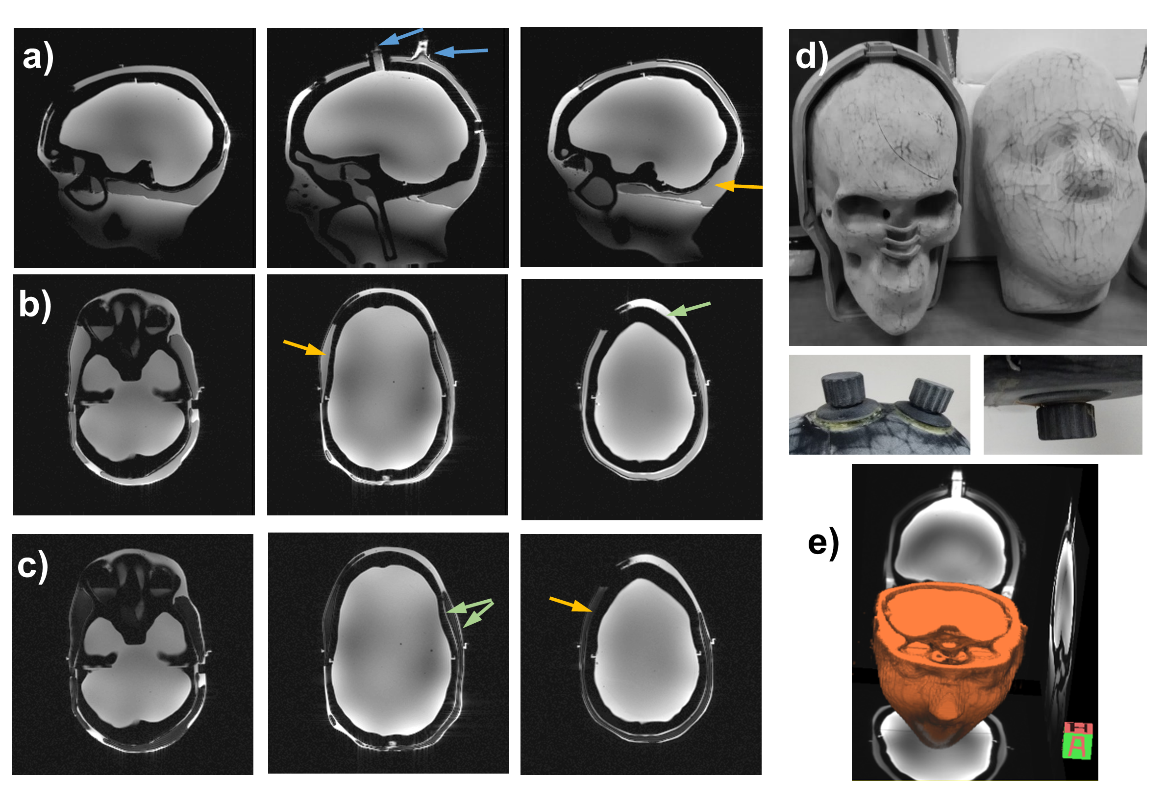
Figure
2: Images of the 3D head-shaped phantom. (a-c) Sagittal and axial GRE images of the
phantom without fat suppression (a and b) and with fat suppression (c).
The blue arrows show the opening for filling. The green arrows show the thin
layer generated around the lipid layer as well as the locally thicker layer of
the “muscle”, which improve the phantom’s resemblance to a realistic brain and
the orange arrows show the location of the “lipid” layer. (d) Photos of the 3D
printed structure (inner and outer halves), two screw caps (left) and bottom cap
(right). (e) 3D rendering of phantom images.
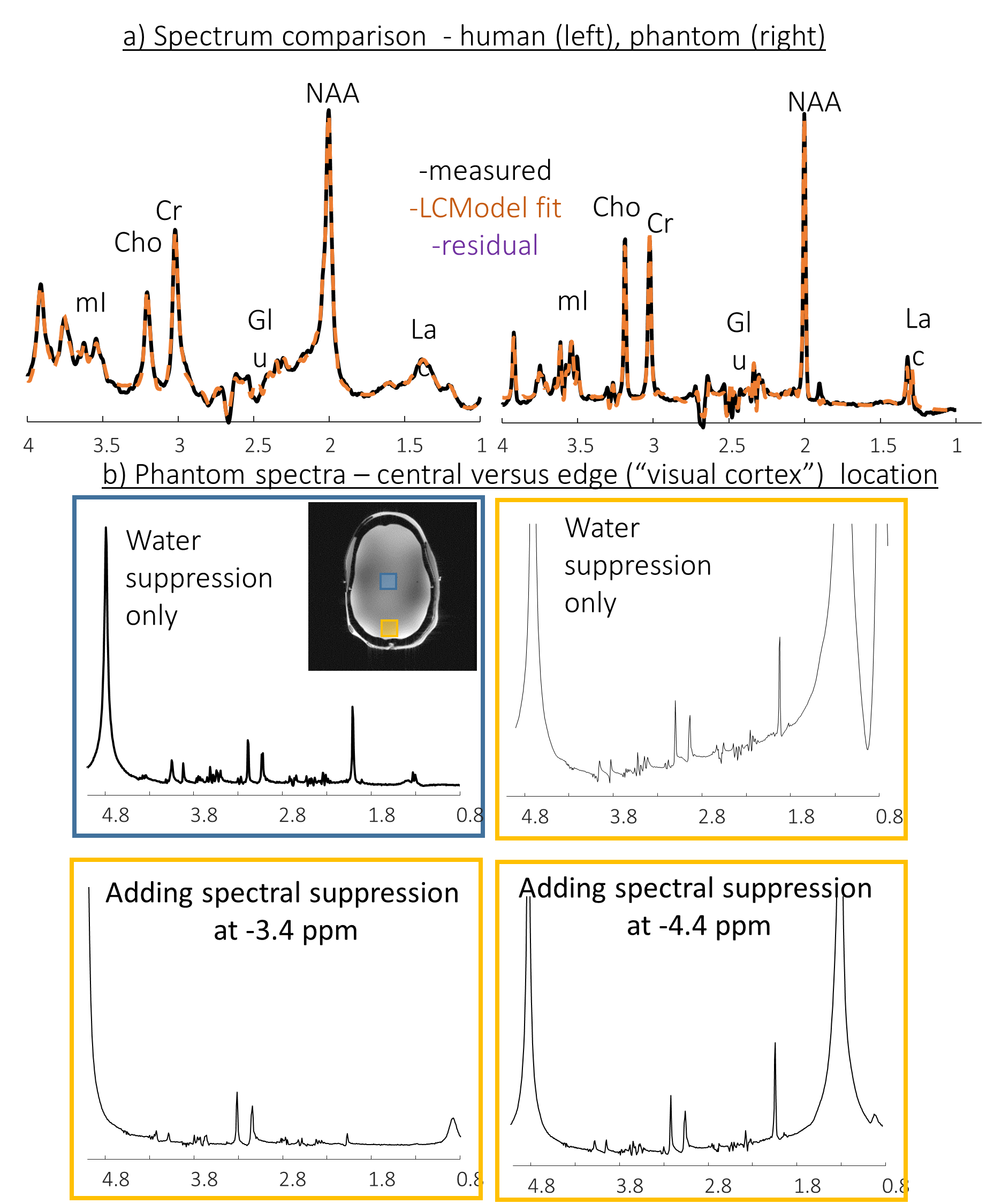
Figure 3: Single voxel spectroscopy. a)
white matter voxel in human (left) and central voxel in the phantom (b) Phantom
spectra – comparison of a central (blue box) and near lipid (“visual cortex”) (orange
box) voxels. The SVS near lipid voxel was repeated without Spectral
Suppression, and with Spectral Suppression at -3.4 ppm and at -4.4 ppm.