Elif Aygun1,2, Ahmet Rahmetullah Cagil1,2, and Emine Ulku Saritas1,2,3
1Department of Electrical and Electronics Engineering, Bilkent University, Ankara, Turkey, 2National Magnetic Resonance Research Center (UMRAM), Bilkent University, Ankara, Turkey, 3Neuroscience Graduate Program, Bilkent University, Ankara, Turkey
1Department of Electrical and Electronics Engineering, Bilkent University, Ankara, Turkey, 2National Magnetic Resonance Research Center (UMRAM), Bilkent University, Ankara, Turkey, 3Neuroscience Graduate Program, Bilkent University, Ankara, Turkey
A 1:1 scale human head and neck agar-agar phantom mimicking human
tissue characteristics is developed using a 3D human model. The phantom closely
matches human anatomy and the susceptibility artifacts in MRI images successfully
mimic those seen during in vivo imaging of the brain and spine.
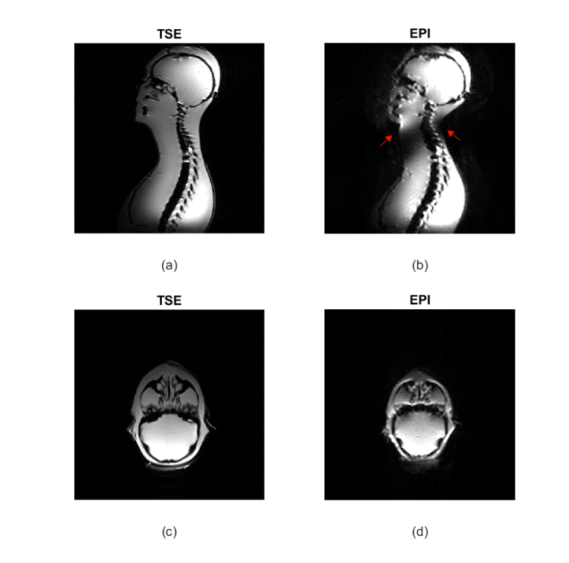
Figure 5. MRI images acquired
with TSE and EPI sequences. (a-b) Sagittal images of the whole phantom and the
spine. The susceptibility artifacts are seen on the back and front of the neck for
EPI (red arrows) successfully mimick the problems seen during in vivo spine
imaging. (c-d) The brain in the axial plane also shows susceptibility artifacts,
similar to the ones seen during in vivo brain imaging.
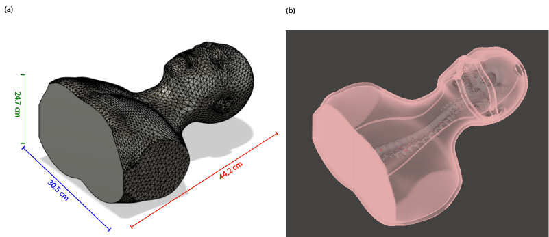
Figure 2. (a) The phantom
dimensions were 30.5 x 24.7 x 44.2 cm3 with 8-mm wall thickness. (b)
The spine and the skull were placed inside of the phantom, aligned post-print with
the center of the phantom. The spine was attached to the skull from the Atlas
bone with plastic cable ties and super glue, and was fixed to the bottom of the
phantom using hot glue. These fixations ensured that the spine remains in its
intended place during the filling of the agar-agar gel.