Emilie Poirion1, Corentine Marie2, Marine Boudot de La Motte3, Chloé Dupont4, Jean-Claude Sadik1, and Julien Savatovsky1
1Hospital Foundation A. de Rothschild, Imaging department, Paris, France, PARIS, France, 2Sorbonne University, Paris Brain Institute, Paris, France, Paris, France, 3Hospital Foundation Rothschild,Neurology department, Paris, France, Paris, France, 4Hospital Foundation A. de Rothschild, Pharmaceutical department, Paris, France, PARIS, France
1Hospital Foundation A. de Rothschild, Imaging department, Paris, France, PARIS, France, 2Sorbonne University, Paris Brain Institute, Paris, France, Paris, France, 3Hospital Foundation Rothschild,Neurology department, Paris, France, Paris, France, 4Hospital Foundation A. de Rothschild, Pharmaceutical department, Paris, France, PARIS, France
To detect low-dose
gadolinium in the meninges, we tested an optimized FLAIR sequence on a custom
gadolinium phantom, showing a higher CNR on this sequence. The first in-vivo
experiment is promising as we observed an improvement of leptomeningeal
enhancement with the optimized sequence.
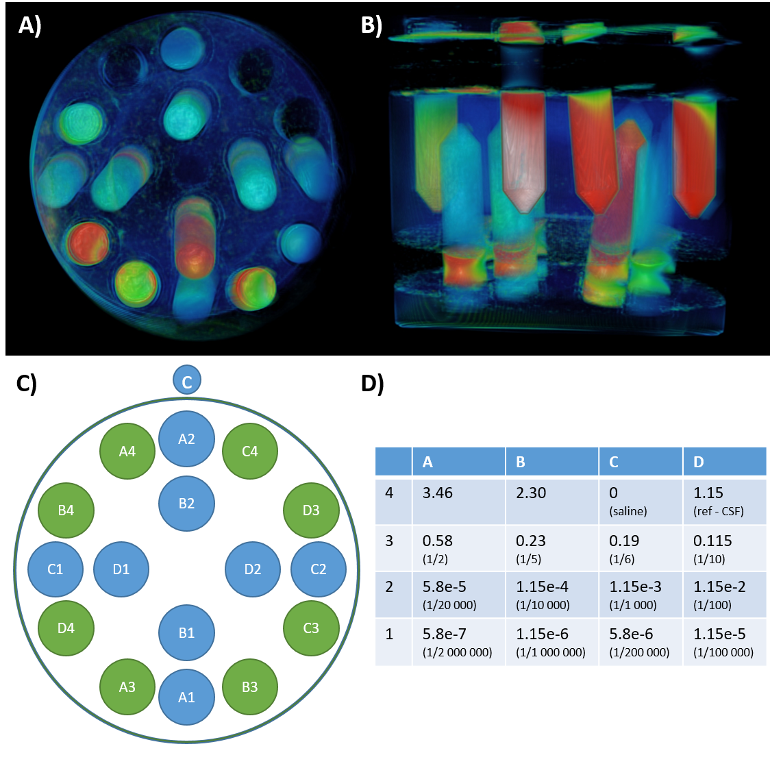
Figure 1: Phantom design.
We prepared a phantom (A, B) composed of 16 tubes (C) with variable concentration of gadolinium (expressed as mg/ml, D), and filled with agar.
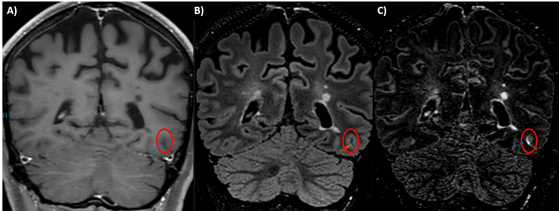
Figure 4: In-vivo preliminary evaluation.
A patient with Multiple Sclerosis (female, 31Y) underwent an MRI as part of her clinical follow-up, which includes a T1-weighted TSE (A), a FLAIR (B), and an optimized FLAIR (C). A linear leptomeningeal enhancement (red circle) was visible on the optimized FLAIR, while totally absent from the T1 TSE, and suspected on the conventional FLAIR.