Egor Kretov1, Zhao Kaixuan 1,2, Charles Grassin3, and Thoralf Niendorf1
1Max Delbrück Center for Molecular Medicine, Berlin, Germany, 2School of Biomedical Engineering, Southern Medical University, Guangzhou, China, 3Independent Researcher, Paris, France
1Max Delbrück Center for Molecular Medicine, Berlin, Germany, 2School of Biomedical Engineering, Southern Medical University, Guangzhou, China, 3Independent Researcher, Paris, France
This work demonstrates a new cost-effective near-field
RF mapping tool that combines computer vision with field measurement techniques
to evaluate MR coils and to study electromagnetic field interferences.
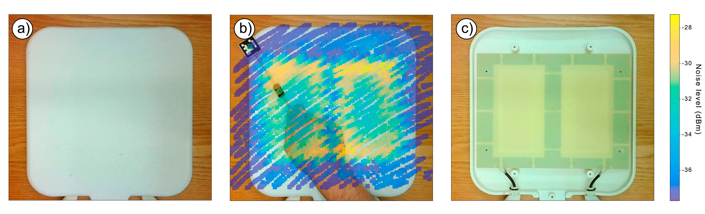
Figure 3. a) Photo of the anterior section of a
7.0 T cardiac RF coil array with hidden internal structure b) manual field
measurements reveal two RF loop elements and with rectangular shape c) picture
photograph of RF coil array with the housing removed confirming the coil
geometry.
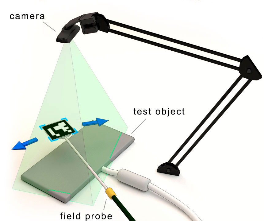
Figure 1. Schematic view of the setup for MR coil
assessment. The camera is positioned over the evaluated object and the green
zone represents the area visible to the camera. The magnetic field probe is
equipped with the QR-code marker from the ArUco library and connected to the
USB SDR receiver.
