Apoorva Agarwal1, Megha Goel1, and Jignesh Dholakia1
1GE Healthcare, Bengaluru, India
1GE Healthcare, Bengaluru, India
A robust anatomy and pulse
sequence-generic technique where 3-plane Localizer data is used to achieve
background-free MR images has been proposed. Better windowing and enhanced visual
contrast along with improved accuracy over previous attempts has been
demonstrated.
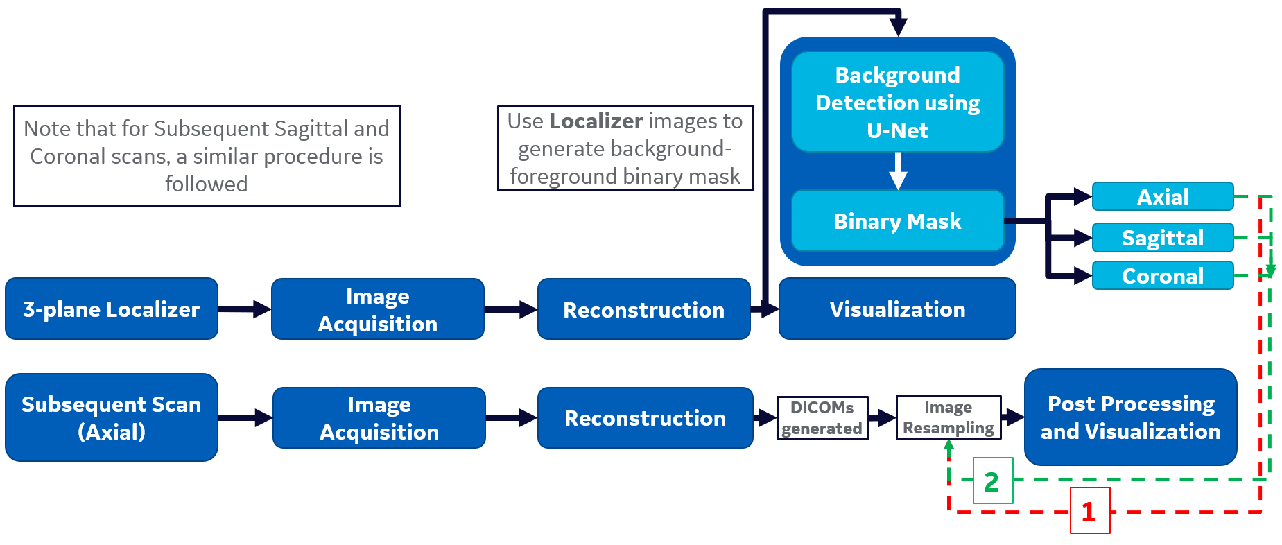
Proposed
workflow – Pipeline 1: Masks from Axial Localizer stack are input to Image Resampling
for background identification in Axial data (follow Red arrow), Pipeline 2: A union of Masks
from Axial, Sagittal and Coronal Localizer stacks is input to Image Resampling for background identification in Axial data
(follow Green arrow).
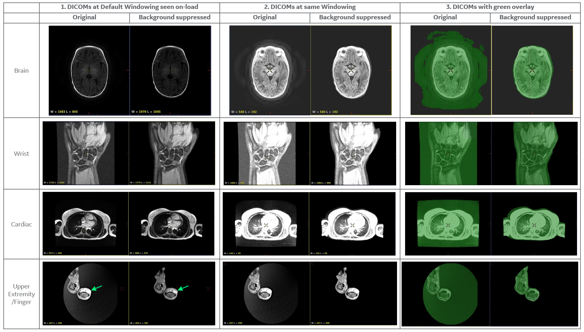
Results of different anatomies obtained using their respective Localizers: Note the change in default WW/WL on-load in AW viewer (Col. 1) (Brain: 1683/845 -> 1970/1045; Wrist: 1734/1042 -> 1775/1114; Cardiac: 871/446 -> 900/475; Finger: 427/250 -> 653/367). The range shifts right and dynamic range of visible pixel intensities increases. Observe better distinction of finger mass boundaries in Row 4. Col. 2 shows results at same, increased windowing to show the interference of background when viewing at higher brightness. Col. 3 shows removed background pixels using green overlay.