Miriam Heynckes1, Elia Formisano1,2, Peter De Weerd1, and Federico De Martino1
1Faculty of Psychology and Neuroscience, Maastricht University, Maastricht, Netherlands, 2Maastricht Centre for Systems Biology, Maastricht University, Maastricht, Netherlands
1Faculty of Psychology and Neuroscience, Maastricht University, Maastricht, Netherlands, 2Maastricht Centre for Systems Biology, Maastricht University, Maastricht, Netherlands
Trial-by-trial layer-specific BOLD responses in primary auditory cortex during temporal deviant detection in rhythmic sounds
Proof of concept of psychophysics task
Spectrally unspecific increase in superficial layers when deviant detected
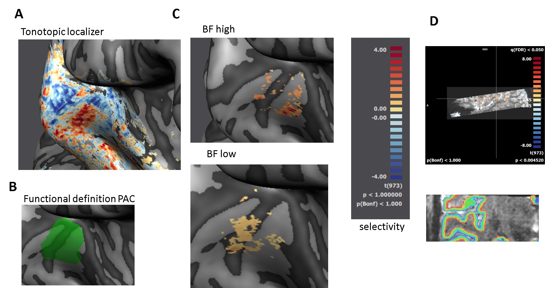
A: Tonotopic map of the right hemisphere (red = low frequency, blue = high frequency). Acquired in a blocked design, at 1.2 mm iso. In total 7 frequencies (130 Hz, 200 Hz, 306 Hz, 721 Hz, 1100
Hz, 1700 Hz and 4000 Hz) were used. B: We functionally defined PAC, following the high-low-high gradient on the medial
portion of Heschl’s gyrus, see (Moerel et al. 2014).
C: Based on the tonotopy, we made selectivity maps to high and low sounds. Difference between center - frequencies is 2.5 octaves. D: Activation map to high and low sounds overlaid on BOLD data.
Equi-volume layers sample the cortical depth.
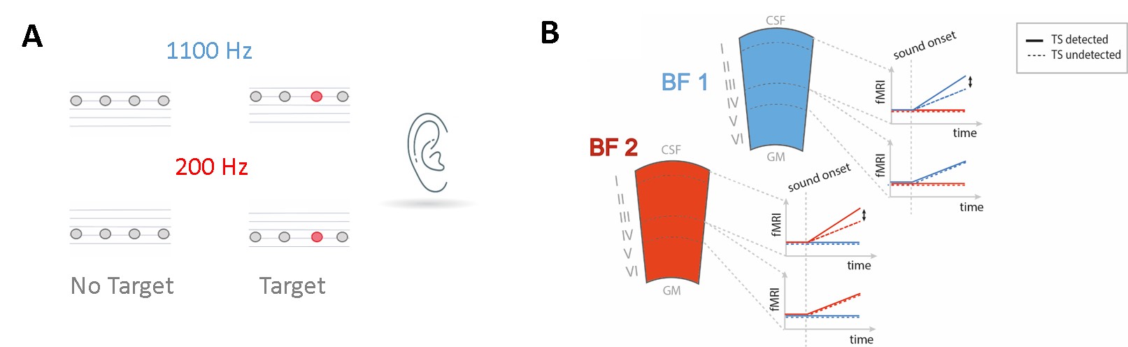
A: Rhythmic 2 Hz sound at a low or high carrier frequency.
Participants detected a temporal deviant (red). Target
difficulty was modulated between 1 -10 ms temporal
shift. B: Hypotheses. Each column depicts a best frequency (BF) region of
PAC with a frequency preference to either BF1 or BF2. Stimuli matching BF
will elicit larger activation in the respective region than non-BF stimuli. An
additional upregulation in superficial layers is expected when detecting a deviant (dotted vs. solid line). We expect this to be
spectrally specific to the BF region matching the stimulus frequency.