Jiajia Yang1,2, Masaki Fukunaga3, Yinghua Yu2,4, Laurentius Huber5, Peter A Bandettini2, and Norihiro Sadato3
1Graduate School of Interdisciplinary Science and Engineering in Health Systems, Okayama University, Okayama, Japan, 2Section on Functional Imaging Methods, National Institute of Mental Health, Bethesda, MD, United States, 3Division of Cerebral Integration, National Institute for Physiological Sciences, Okazaki, Japan, 4Center for Information and Neural Networks, National Institute of Information and Communications Technology, Suita, Japan, 5Department of Cognitive Neuroscience, Maastricht University, Maastricht, Netherlands
1Graduate School of Interdisciplinary Science and Engineering in Health Systems, Okayama University, Okayama, Japan, 2Section on Functional Imaging Methods, National Institute of Mental Health, Bethesda, MD, United States, 3Division of Cerebral Integration, National Institute for Physiological Sciences, Okazaki, Japan, 4Center for Information and Neural Networks, National Institute of Information and Communications Technology, Suita, Japan, 5Department of Cognitive Neuroscience, Maastricht University, Maastricht, Netherlands
We found the prediction relative double-peak activity feature across midcingulate cortex layers using high-resolution BOLD and VASO fMRI at 7T. We regard the finding as an essential step towards the understanding of predictive coding processing.
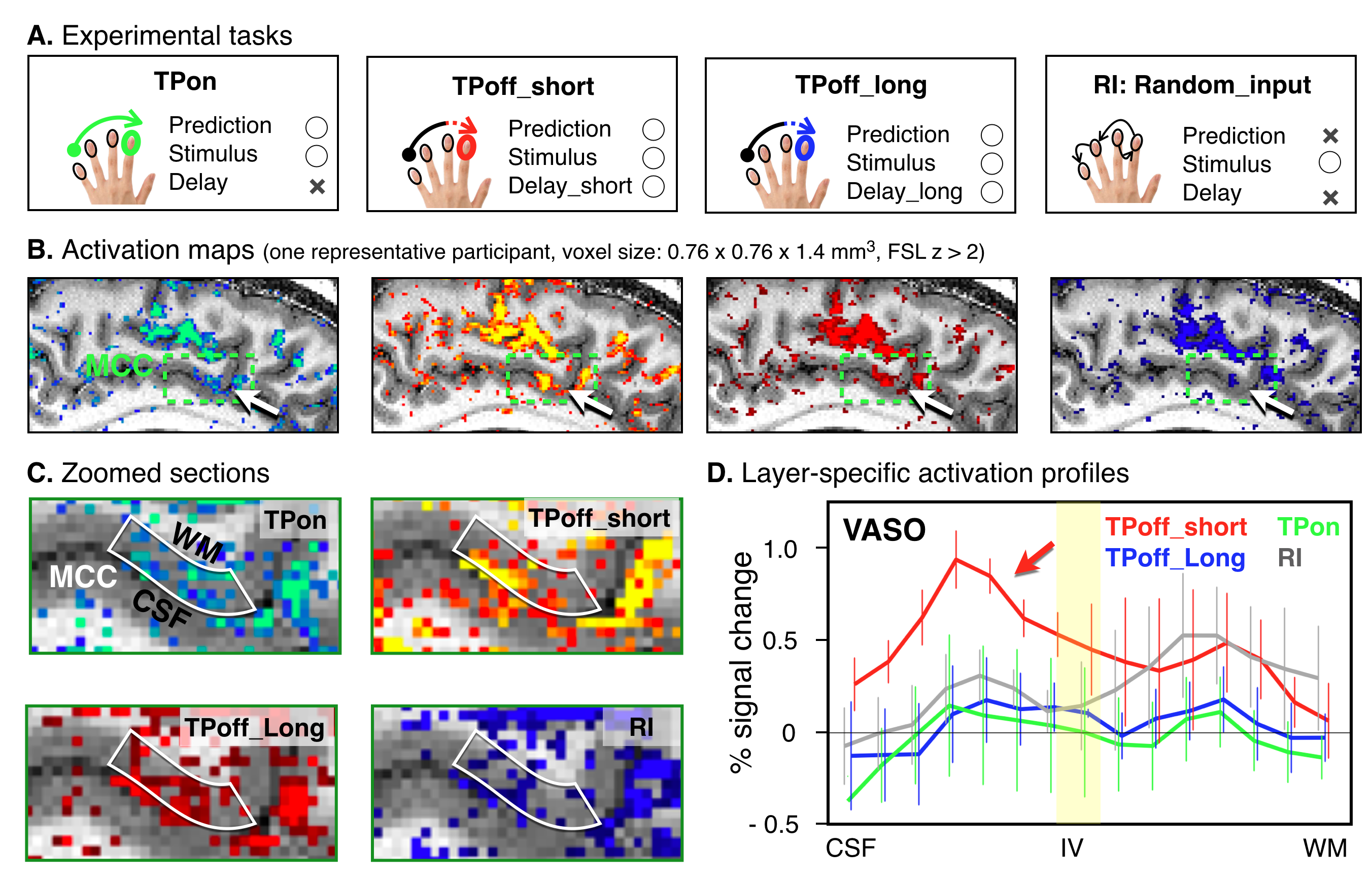
Figure 2. (A) The four experimental tasks are illustrated.(B) Smoothed BOLD activation maps of four different tasks of one participant are shown. (C) Zoomed MCC sections of unsmoothed activation maps are shown.It can be seen that the four tasks have a different distribution of activity across the layers in MCC. (D) Averaged (n=4) cortical profiles of VASO activity changes in MCC are shown.We found the double-peak activity feature across MCC layers for all tasks. The TPoff_short task (red line) enhanced activity within the superficial layers than all other tasks.
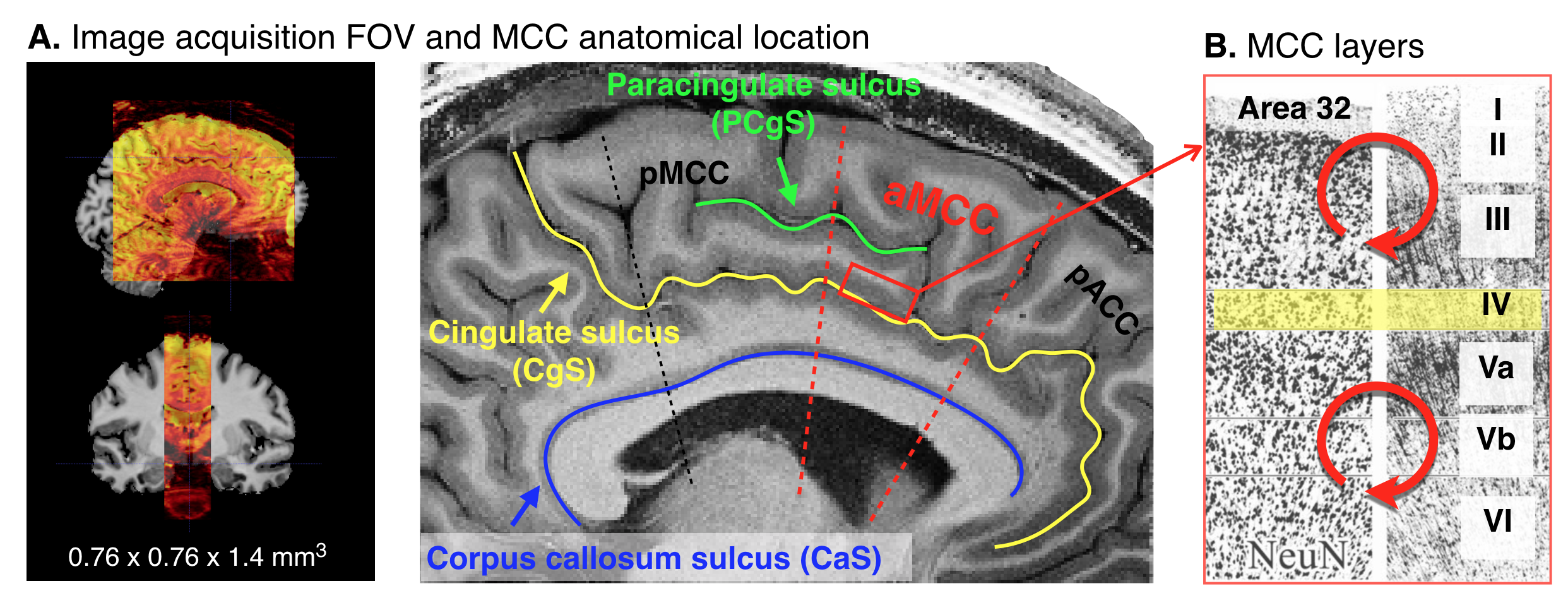
Figure 1. (A) The image acquisition FOV and MCC anatomical location are shown. Slice-selective slab-inversion VASO was used on a 7T scanner, equipped with a 32-channel RF coil and a SC72 body gradient coil. The nominal resolution was 0.76 mm across cortical depths with 1.4-mm thick slices. (B) Illustration of MCC layers.