Michela Pievani1, Ileana O. Jelescu2, Joao Jorge3, Olivier Reynaud4, Federica Ribaldi1,5,6, Valentina Garibotto7, Giovanni B. Frisoni1,5, and Jorge Jovicich8
1Laboratory of Alzheimer’s Neuroimaging and Epidemiology, IRCCS Istituto Centro San Giovanni di Dio Fatebenefratelli, Brescia, Italy, 2CIBM - Center for Biomedical Imaging, Ecole Polytechnique Fédérale de Lausanne, Lausanne, Switzerland, 3Systems Division, Swiss Center for Electronics and Microtechnology (CSEM), Nêuchatel, Switzerland, 4Human Neuroscience Platform, Fondation Campus Biotech Geneva, Geneva, Switzerland, 5Memory Clinic and LANVIE - Laboratory of Neuroimaging of Aging, University Hospitals and University of Geneva, Geneva, Switzerland, 6Department of Molecular and Translational Medicine, University of Brescia, Brescia, Italy, 7Division of Nuclear Medicine and NIMTlab, University Hospitals and University of Geneva, Geneva, Switzerland, 8Center for Mind/Brain Sciences, University of Trento, Trento, Italy
1Laboratory of Alzheimer’s Neuroimaging and Epidemiology, IRCCS Istituto Centro San Giovanni di Dio Fatebenefratelli, Brescia, Italy, 2CIBM - Center for Biomedical Imaging, Ecole Polytechnique Fédérale de Lausanne, Lausanne, Switzerland, 3Systems Division, Swiss Center for Electronics and Microtechnology (CSEM), Nêuchatel, Switzerland, 4Human Neuroscience Platform, Fondation Campus Biotech Geneva, Geneva, Switzerland, 5Memory Clinic and LANVIE - Laboratory of Neuroimaging of Aging, University Hospitals and University of Geneva, Geneva, Switzerland, 6Department of Molecular and Translational Medicine, University of Brescia, Brescia, Italy, 7Division of Nuclear Medicine and NIMTlab, University Hospitals and University of Geneva, Geneva, Switzerland, 8Center for Mind/Brain Sciences, University of Trento, Trento, Italy
Functional
connectivity (FC) of the human locus coeruleus at 7T is positive with
the cerebellum and the frontal cortex. The default mode and
frontoparietal networks, but not the salience network, show FC with
the brainstem.
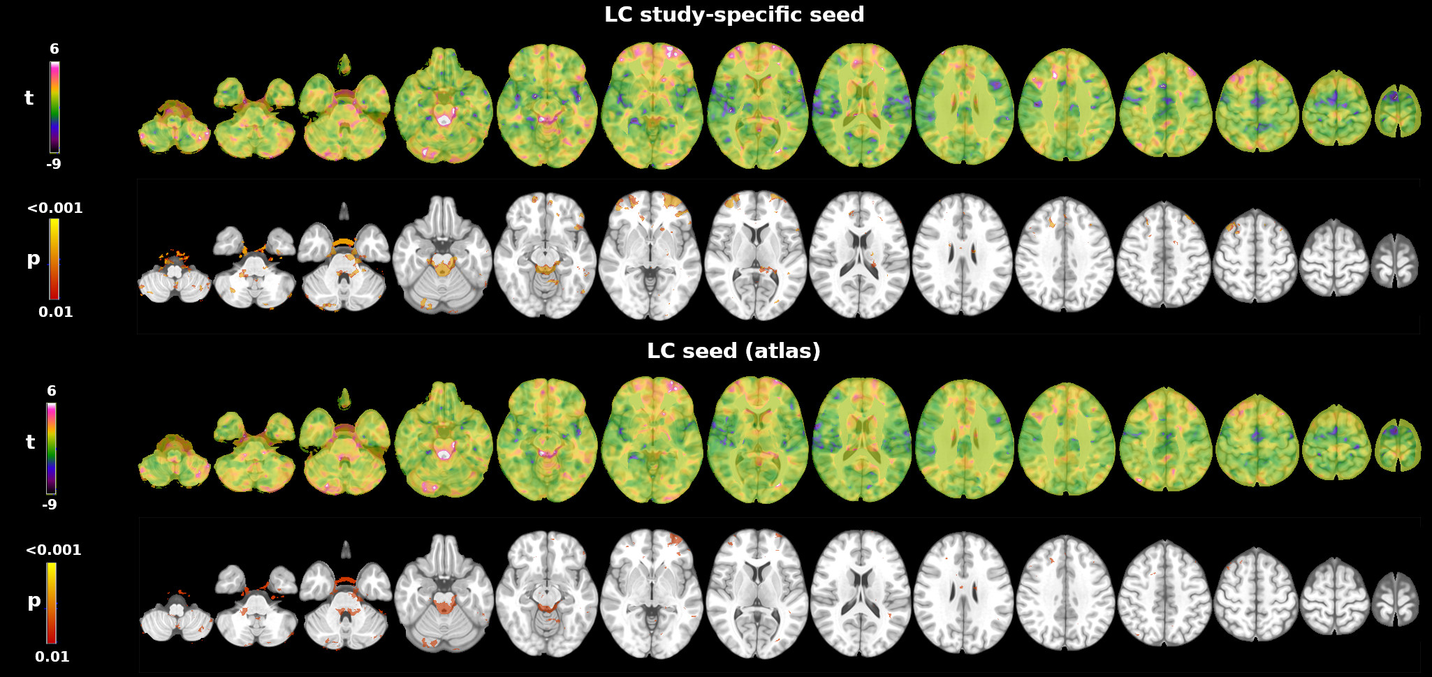
Figure
1. Whole
brain 7T resting-state functional connectivity of the locus
coeruleus (LC).
Top: LC
functional connectivity map using the study specific LC as seed.
Bottom: LC connectivity maps using a published11 7T LC atlas as seed.
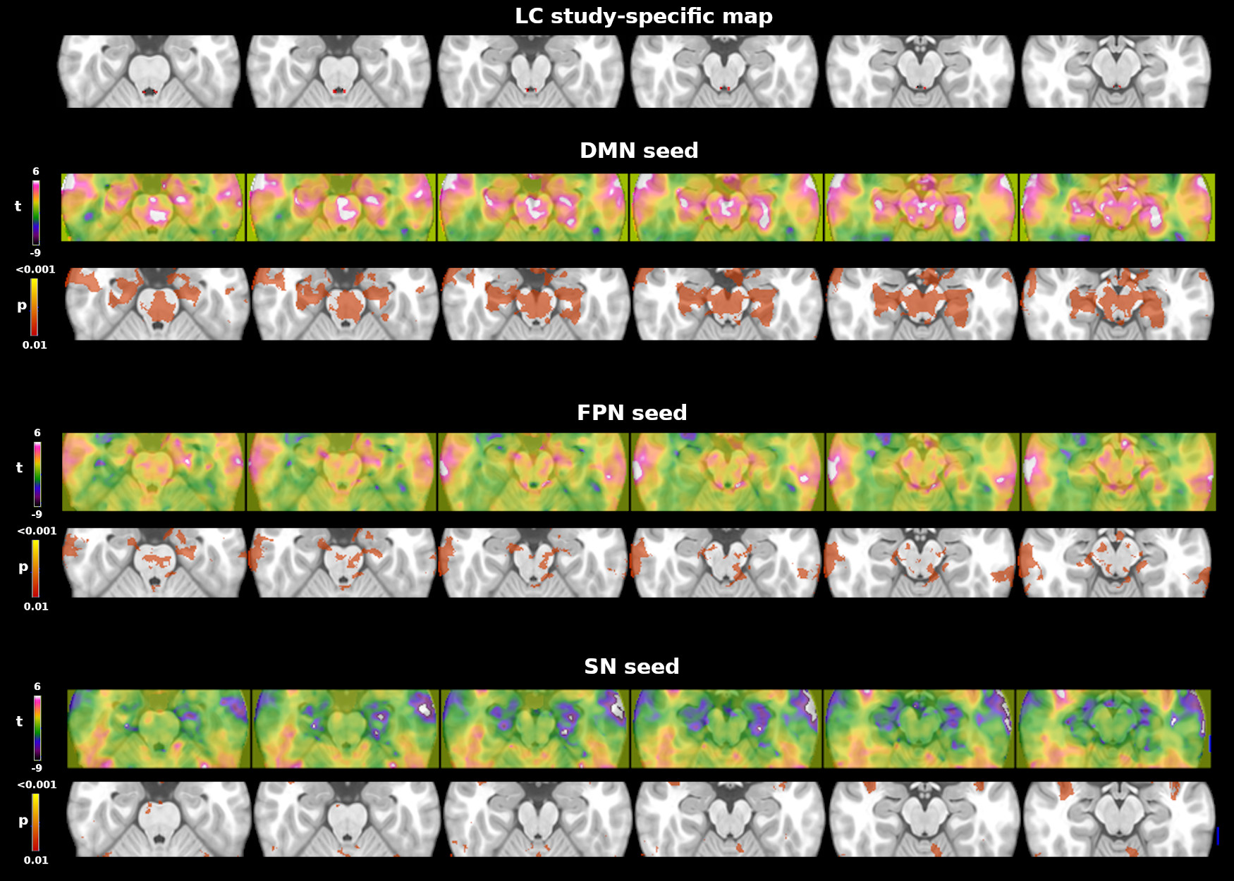
Figure
2.
Resting-state
functional connectivity map
using the default mode network (DMN; top), fronto parietal network
(FPN; middle) and salience network (SN, bottom) atlas12 as seed. The
figure shows brainstem areas functionally connected with the seed.
The LC spatial map (top) is provided for anatomical reference.