Jonathan R. Polimeni1,2, Olivia M. Viessmann1, Qiyuan Tian1, Michaël Bernier1, Meher R. Juttukonda1, Yi-Fen Yen1, and David H. Salat1,3
1Athinoula A. Martinos Center for Biomedical Imaging, Massachusetts General Hospital/Harvard Medical School, Charlestown, MA, United States, 2Division of Health Sciences and Technology, Massachusetts Institute of Technology, Cambridge, MA, United States, 3Neuroimaging Research for Veterans Center, VA Boston Healthcare System, Boston, MA, United States
1Athinoula A. Martinos Center for Biomedical Imaging, Massachusetts General Hospital/Harvard Medical School, Charlestown, MA, United States, 2Division of Health Sciences and Technology, Massachusetts Institute of Technology, Cambridge, MA, United States, 3Neuroimaging Research for Veterans Center, VA Boston Healthcare System, Boston, MA, United States
t.b.d.
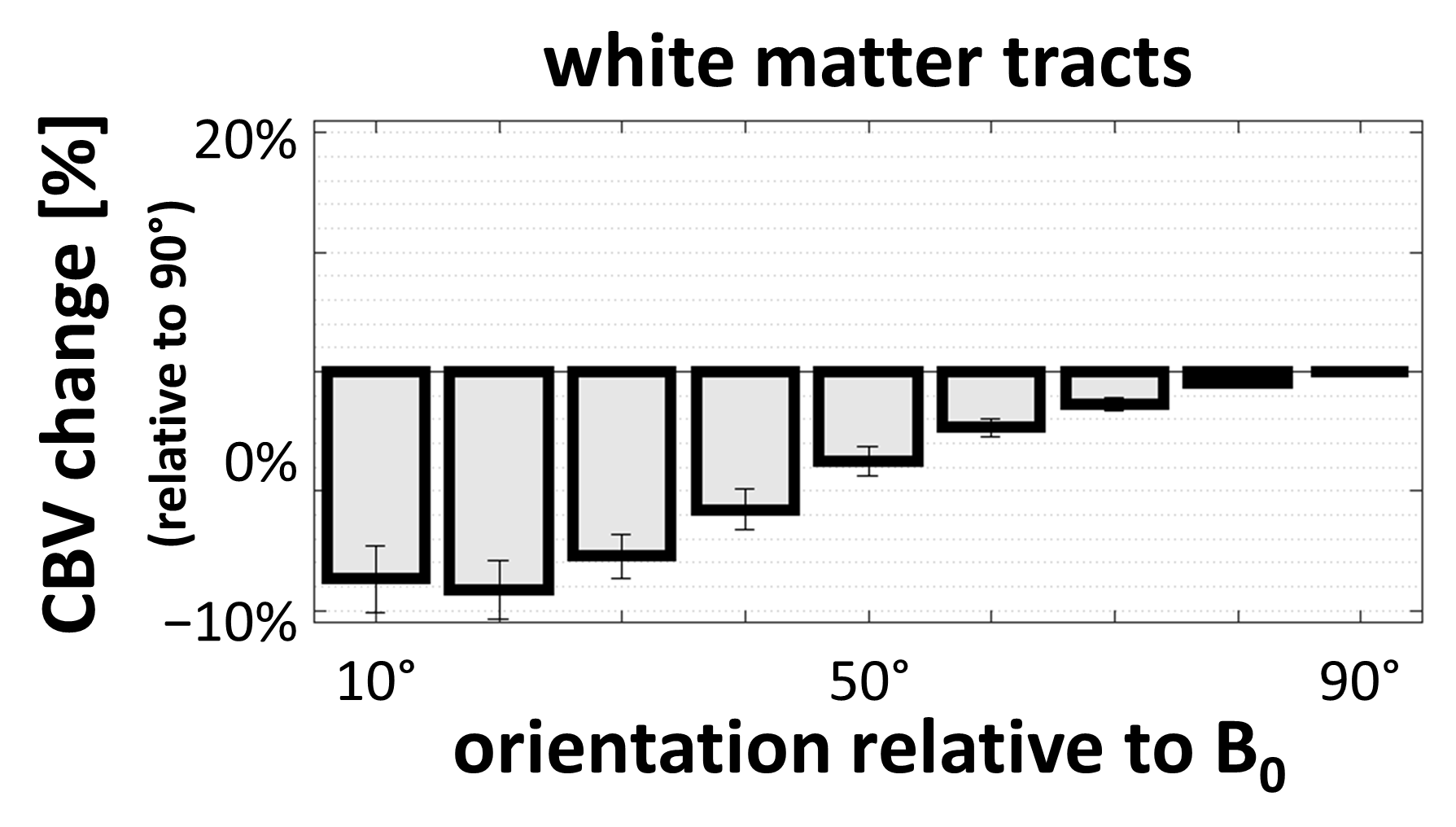
Figure 5: White matter fiber tract orientation dependence of
the baseline CBV estimates, averaged across subjects. Consistently lower CBV values are observed in
tracts where the tract orientation is parallel to the B0 direction. This is
consistent with the largest blood vessels contributing to the DSC-based
estimates of CBV in the white matter running parallel to the white matter
tracts derived from these diffusion data. (Error bars indicate standard error across subjects.)
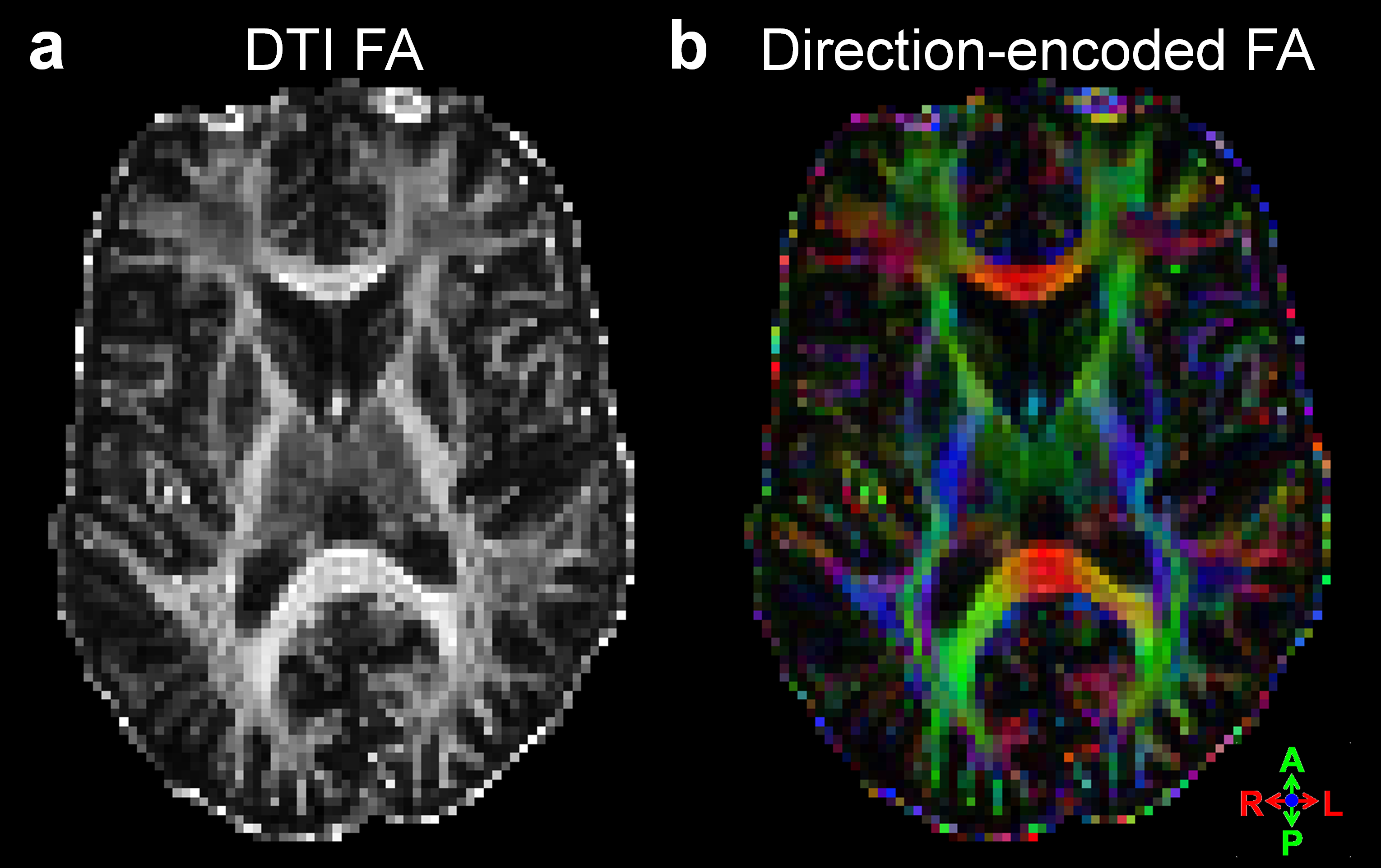
Figure 2: Example 7T diffusion data and associated fractional anisotropy maps presented as (a) gray-scale and (b) direction-encoded color-scale.