An Thanh Vu1,2, Alexander JS Beckett3,4, Jennifer Townsend3,4, Salvatore Torrisi3,4, and David Feinberg3,4
1Radiology, University of California, San Francisco, San Francisco, CA, United States, 2San Francisco Veteran Affairs Health Care System, San Francisco, CA, United States, 3Advanced MRI Technologies, Sebastopol, CA, United States, 4Helen Wills Neuroscience Institute, University of California, Berkeley, Berkeley, CA, United States
1Radiology, University of California, San Francisco, San Francisco, CA, United States, 2San Francisco Veteran Affairs Health Care System, San Francisco, CA, United States, 3Advanced MRI Technologies, Sebastopol, CA, United States, 4Helen Wills Neuroscience Institute, University of California, Berkeley, Berkeley, CA, United States
In this 7T study, we found that through constructive combination of the BOLD and CBV weighted timeseries of EPI and GRASE based Vascular Space Occupancy (VASO) sequences fMRI CNR could be significantly enhanced relative to BOLD alone.
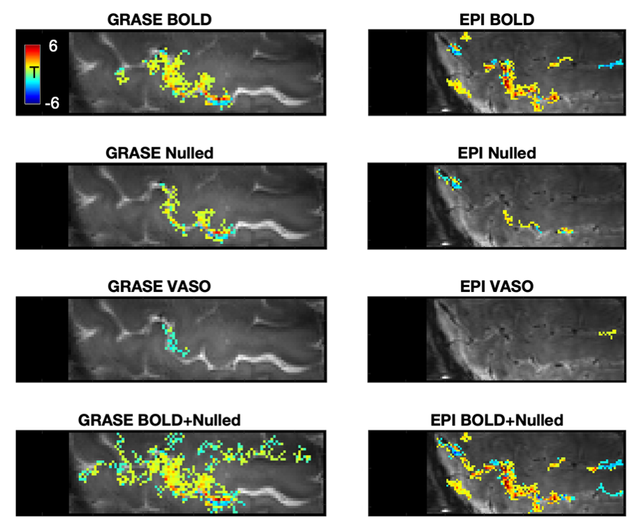
Figure 1. A representative slice from one subject in terms of fMRI CNR and extent of activation (above-threshold t-values overlaid onto the timeseries mean BOLD image) for GRASE (left) and EPI (right) versions of the VASO sequence. The BOLD+Blood Nulled combination (bottom) compared to BOLD alone (top), shows stronger fCNR with broader spatial extent of activations.
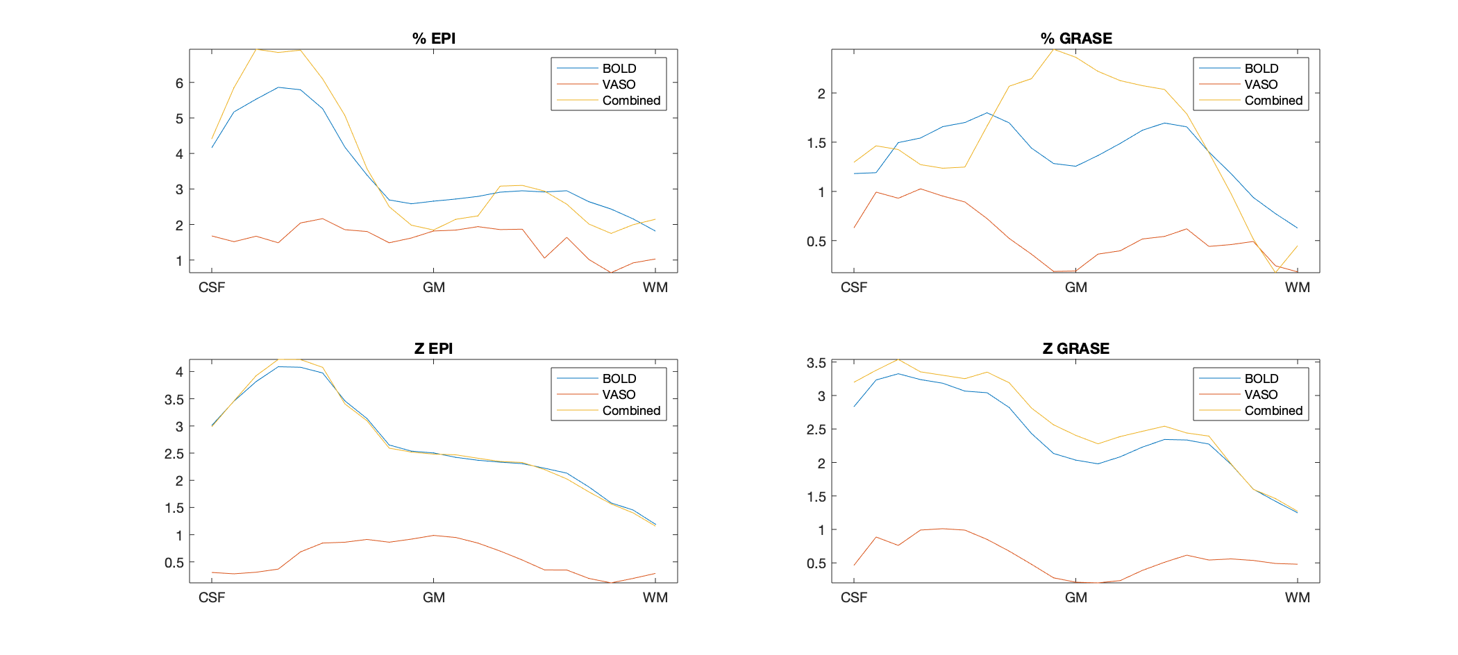
Figure 5. Average motor cortex depth profiles, averaged across subjects, for signal change between superficial (toward CSF) and deep (towards WM) depths for Percent (%) Signal Change (top) and task activation Z-Score (bottom).