Seong Dae Dae Yun1, Patricia Pais-Roldán1, Nicola Palomero-Gallagher2,3,4, and N. Jon Shah1,5,6,7
1Institute of Neuroscience and Medicine 4, INM-4, Forschungszentrum Juelich, Juelich, Germany, 2Institute of Neuroscience and Medicine 1, INM-1, Forschungszentrum Juelich, Juelich, Germany, 3C. & O. Vogt Institute for Brain Research, Heinrich-Heine-University, Duesseldorf, Germany, 4Department of Psychiatry, Psychotherapy and Psychosomatics, Medical Faculty, RWTH Aachen, Aachen, Germany, 5Institute of Neuroscience and Medicine 11, INM-11, JARA, Forschungszentrum Juelich, Juelich, Germany, 6JARA - BRAIN - Translational Medicine, Aachen, Germany, 7Department of Neurology, RWTH Aachen University, Aachen, Germany
1Institute of Neuroscience and Medicine 4, INM-4, Forschungszentrum Juelich, Juelich, Germany, 2Institute of Neuroscience and Medicine 1, INM-1, Forschungszentrum Juelich, Juelich, Germany, 3C. & O. Vogt Institute for Brain Research, Heinrich-Heine-University, Duesseldorf, Germany, 4Department of Psychiatry, Psychotherapy and Psychosomatics, Medical Faculty, RWTH Aachen, Aachen, Germany, 5Institute of Neuroscience and Medicine 11, INM-11, JARA, Forschungszentrum Juelich, Juelich, Germany, 6JARA - BRAIN - Translational Medicine, Aachen, Germany, 7Department of Neurology, RWTH Aachen University, Aachen, Germany
TR-external EPIK enabled the identification of
resting-state networks distributed throughout the brain, with half-millimetre
in-plane resolution at 7T. Mapping of resting-state networks with the spatial
resolution and brain coverage provided here has not been previously achieved.
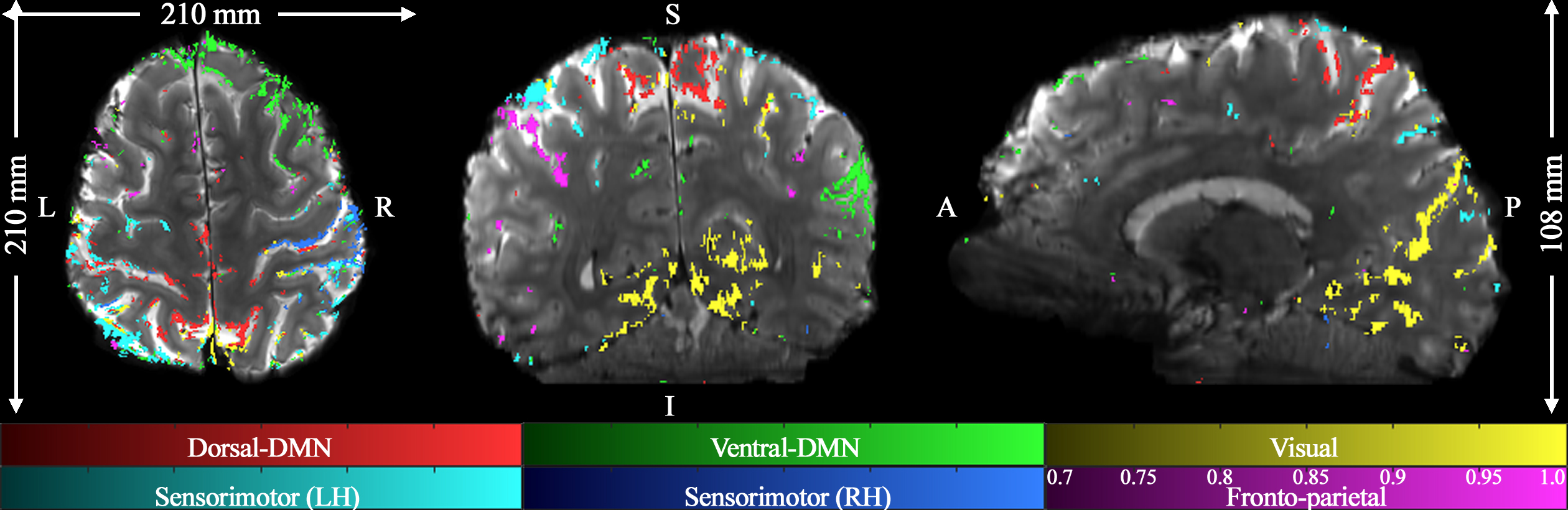
Figure 2. Mapping of six
representative resting-state networks with the half-millimetre protocol (0.51 ×
0.51 × 1.0 mm3) is shown: dorsal-DMN, ventral-DMN, visual,
sensorimotor (LH: Left Hemisphere), sensorimotor (RH: Right Hemisphere) and
fronto-parietal; DMN denote the default mode network. The results, depicted in
three sectional views (axial, coronal and sagittal) above, effectively
demonstrate the brain coverage provided by TR-external EPIK (210 × 210 × 108 mm3)
as well as the localisation of the functional voxels on the cortical ribbon.
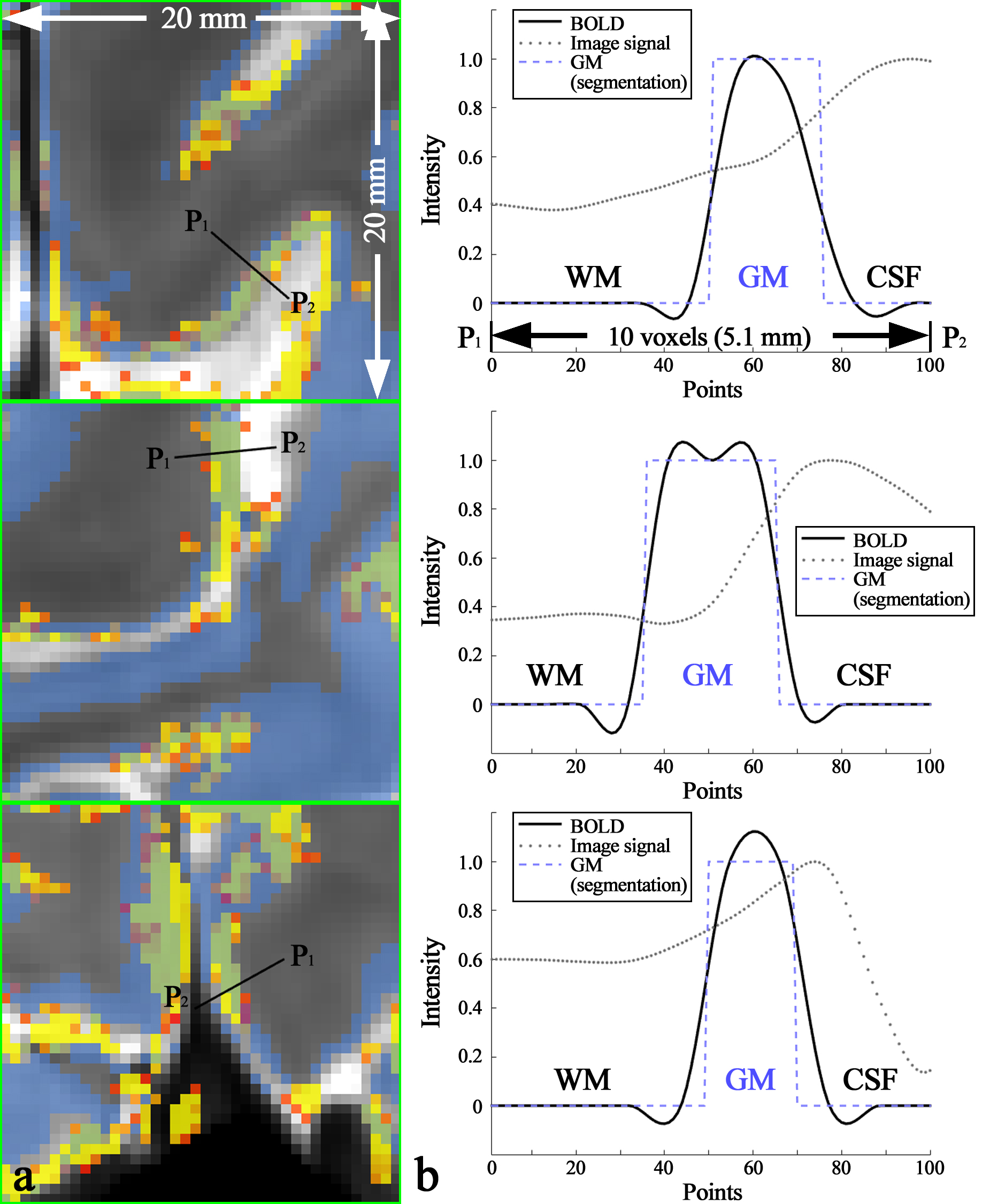
Figure 4. (a) Enlarged depiction of the ROIs marked by the green rectangles in
the right column of Fig. 3 and (b)
line profile of BOLD signals sampled along the black line, denoted with the
points P1 (start) and P2 (end) in the image panel a. The
examined line length was 5.1 mm (10 voxels) and 100 points are equidistantly
re-sampled on this line. The image panel b demonstrates that the resting-state
activation was characterised along the cortical depth for the three networks (dorsal-DMN,
sensorimotor (RH) and visual) and that they are almost confined to the GM
region.