Luca Vizioli1,2, Logan T Dowdle1, Steen Moeller1, Essa Yacoub1, and Kamil Ugurbil1
1CMRR, University of Minnesota, minneapolis, MN, United States, 2Department of Neurosurgery, University Of Minnesota, minneapolis, MN, United States
1CMRR, University of Minnesota, minneapolis, MN, United States, 2Department of Neurosurgery, University Of Minnesota, minneapolis, MN, United States
Using the recently developed NORDIC denoising, we have for the first time achieved robust, 7T gradient echo functional mapping at 0.5mm isotropic in a short experimental time frame. Preliminary layer findings suggest that this may dramatically improve depth-dependent activation profiles.
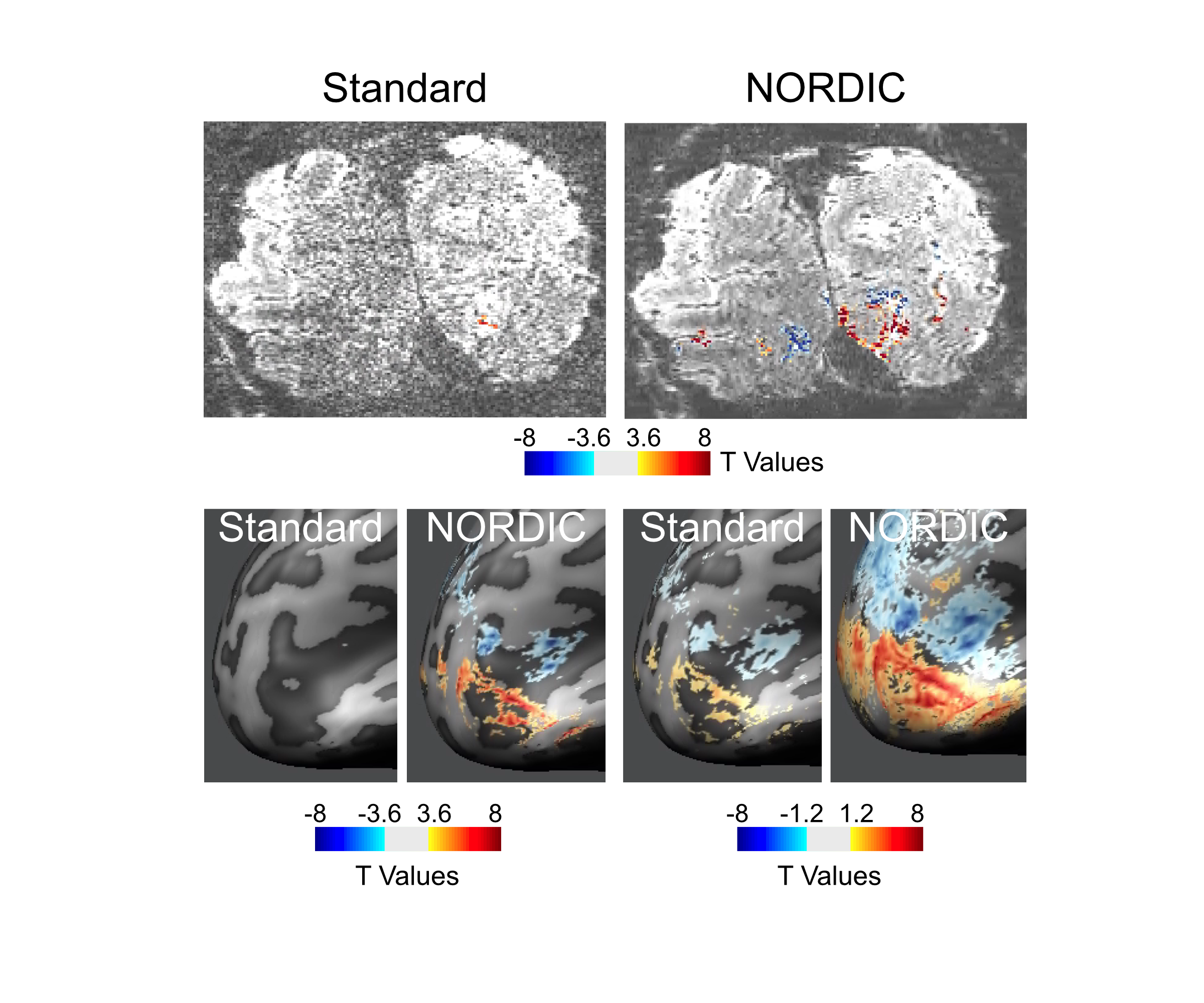
Activation maps (t-values). Top: Examples of NORDIC and Standard reconstructed t-maps superimposed onto a single epi slice obtained from 4 runs (~ 20 minutes of data). The t-maps were computed by contrasting the activation elicited by the target (red) versus that elicited by the surround (blue) condition. Maps were thresholded with t ≥ 3.6 (corresponding to p<.05 FDR corrected for the Standard reconstruction) Bottom: the same t-maps on inflated brains shown at 2 different thresholds, specifically, t ≥ 3.6 (left) and t ≥ 1.2 (right).
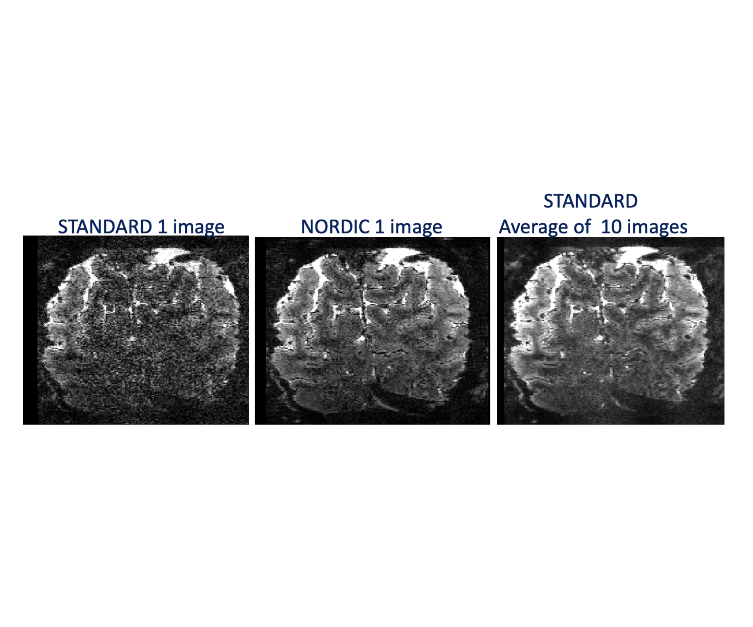
EPI Images. Image of selected single slice from the Standard reconstruction (left), the same single slice from NORDIC reconstructed data (middle), and the average of 10 images of the same slice for the Standard reconstruction (right).