Hankyeol Lee1, Seong-Gi Kim1,2, and Kâmil Uludağ1,2,3
1Center for Neuroscience Imaging Research, Institute for Basic Science, Suwon, Korea, Republic of, 2Department of Biomedical Engineering, Sungkyunkwan University, Suwon, Korea, Republic of, 3Techna Institute & Koerner Scientist in MR Imaging, University Health Network, Toronto, ON, Canada
1Center for Neuroscience Imaging Research, Institute for Basic Science, Suwon, Korea, Republic of, 2Department of Biomedical Engineering, Sungkyunkwan University, Suwon, Korea, Republic of, 3Techna Institute & Koerner Scientist in MR Imaging, University Health Network, Toronto, ON, Canada
Black colored visual stimuli
induced greater BOLD responses in human V1 compared to white stimuli. Voxel
centroid mapping was introduced and used to allocate relative cortical depth
values to all voxels in activation ROIs in multi-subject analysis.
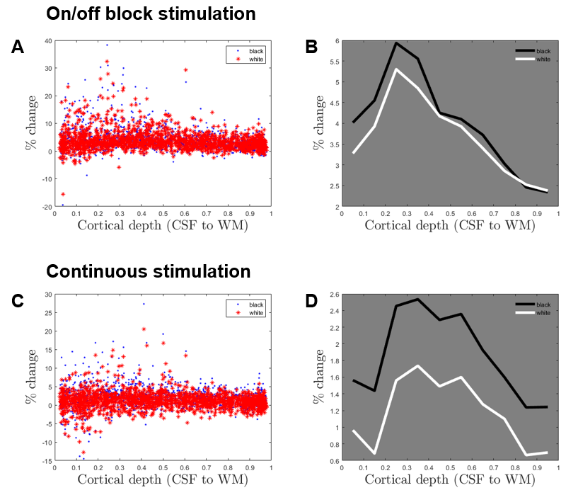
Figure
5. BOLD activation in the masked V1 gray matter voxels plotted against their
cortical depths. A and C show concatenated percent change values for all masked
voxels in 6 subjects (averaged per session). B and D show averaged activation
per cortical depth bins, 10 of them linearly placed along the normalized depth.
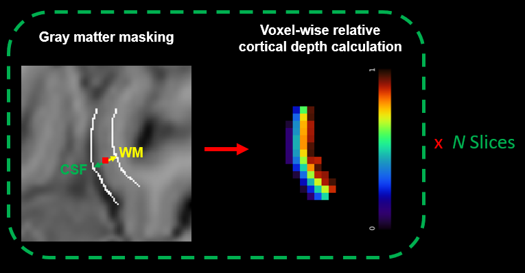
Figure 2. Illustration
of estimating voxel-wise cortical depth using the centroid mapping method. Gray
matter masks are drawn along with borders in an upsampled resolution.
Subsequently, each voxel’s relative distance to the borders (white matter and
CSF) is estimated and its normalized relative cortical depth is assigned.