Marta Xavier1, Inês Esteves1, Athanasios Vourvopoulos1, Ana R Fouto1, Amparo Ruiz-Tagle1, Raquel Gil-Gouveia2, and Patrícia Figueiredo1
1Department of Bioengineering, Institute for Systems and Robotics, Lisbon, Portugal, 2Neurology Department, Hospital da Luz, Lisbon, Portugal
1Department of Bioengineering, Institute for Systems and Robotics, Lisbon, Portugal, 2Neurology Department, Hospital da Luz, Lisbon, Portugal
We demonstrated the viability of the proposed EEG models in predicting the simultaneous fMRI signal from the DMN. We showed that measures of similarity between the predicted and target BOLD signal significantly varied with the model used.
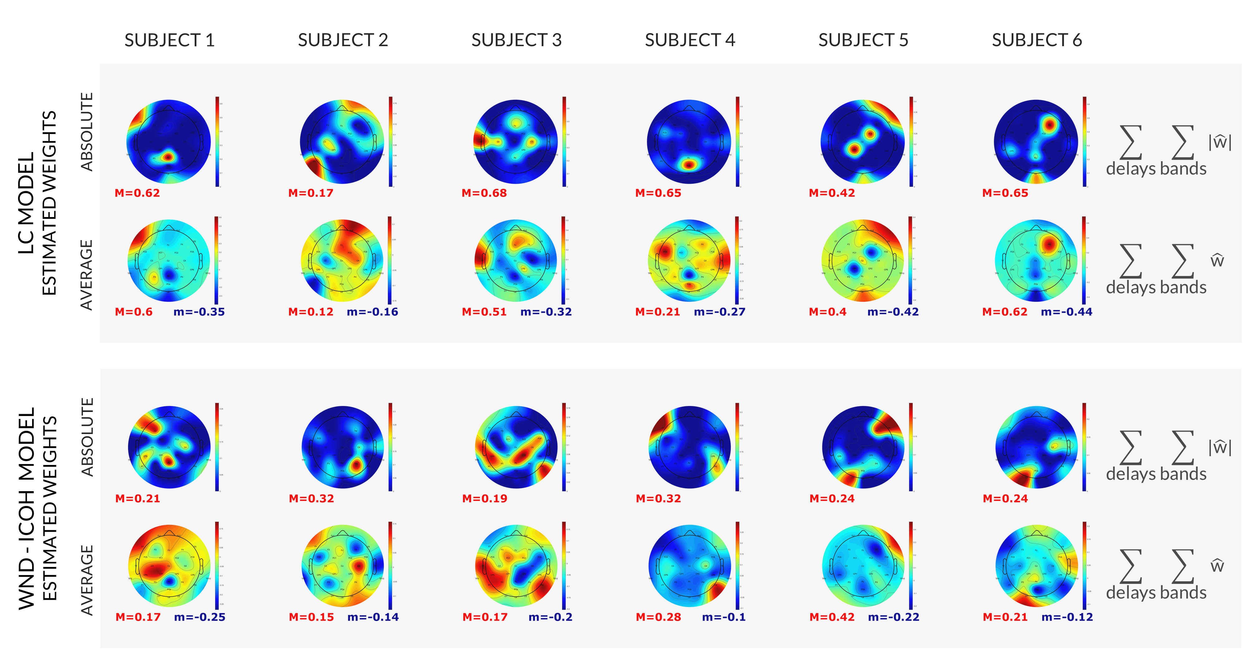
Figure 3: Topographic representation of the estimated model weights, obtained independently for each subject (columns), for the models LC and WD-Icoh (rows). Bottom: estimated weights (w), summed across frequency bands and delays. Top: absolute values of the estimated weights (|w|), summed across frequency bands delays. Minimum and maximum values indicated for each map with m (blue) and M (red).
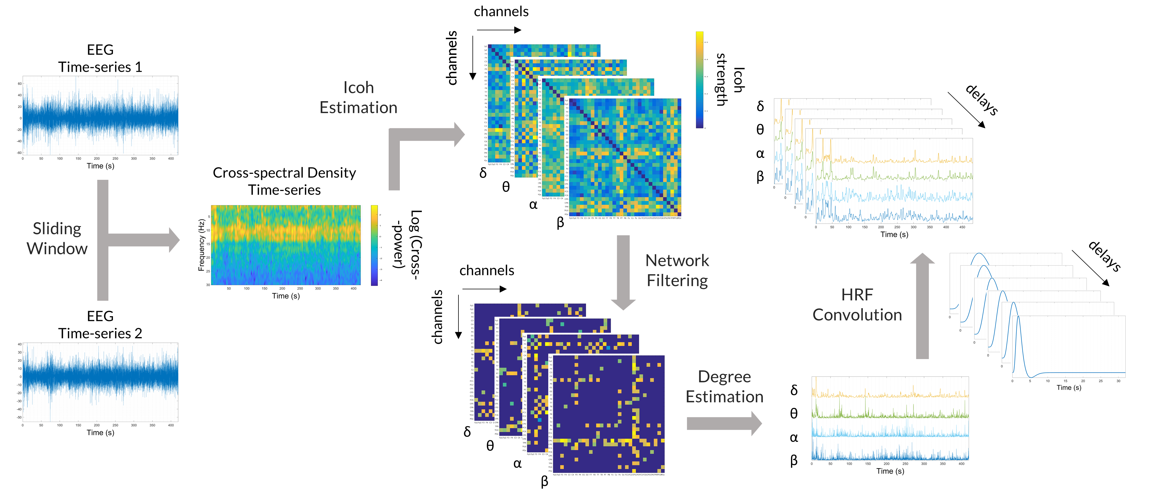
Figure 2: Extraction of the features for the WD-Icoh model. The cross-spectrum was estimated for a hundred frequency values (1-30Hz) in a sliding window of 4s (250ms step size). In each window, the expected value of the cross-spectral density was estimated using Welch's periodogram method. The imaginary part of coherency (Icoh) between each channel pair was estimated and filtered for significance. The weighted degree of each channel was then estimated and averaged for the δ, θ, α and β band.