Rodolfo Abreu1, Júlia F. Soares1, Sónia Batista2,3, Lívia Sousa2,3, Miguel Castelo-Branco1,3, and João Valente Duarte1,3
1Coimbra Institute for Biomedical Imaging and Translational Research (CIBIT), Institute for Nuclear Sciences Applied to Health (ICNAS), University of Coimbra, Coimbra, Portugal, 2Neurology Department, Centro Hospitalar e Universitário de Coimbra, Coimbra, Portugal, 3Faculty of Medicine, University of Coimbra, Coimbra, Portugal
1Coimbra Institute for Biomedical Imaging and Translational Research (CIBIT), Institute for Nuclear Sciences Applied to Health (ICNAS), University of Coimbra, Coimbra, Portugal, 2Neurology Department, Centro Hospitalar e Universitário de Coimbra, Coimbra, Portugal, 3Faculty of Medicine, University of Coimbra, Coimbra, Portugal
Our comparison
revealed that combining a beamformer for EEG source reconstruction with
fMRI-derived spatial priors improves the quality of the reconstructions in
terms of the overlap with atlas-based anatomical brain regions known
to be involved in similar tasks, and RSN templates.
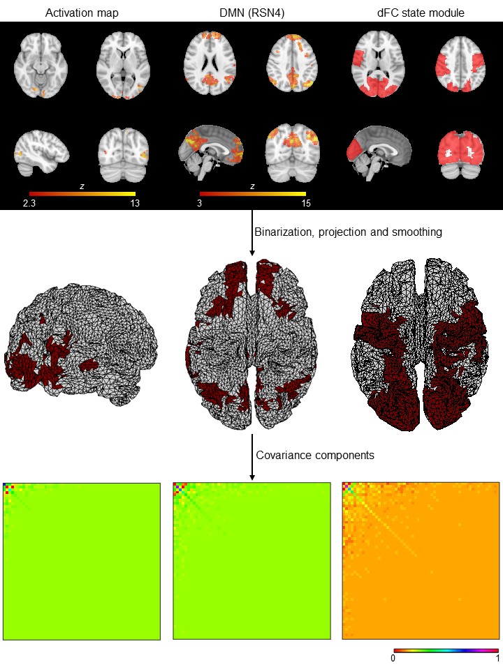
Fig. 2: Deriving covariance components (CCs) from fMRI spatial priors. The
3D fMRI spatial priors are first binarized, projected onto the 2D cortical
surface using nearest-neighbor interpolation and smoothed using the Green’s
function. The associated CCs are then obtained by computing the outer product.
For visualization purposes, the temporally reduced CCs are illustrated, by
applying the same temporal projector considered when reducing the EEG data
prior to its reconstruction.
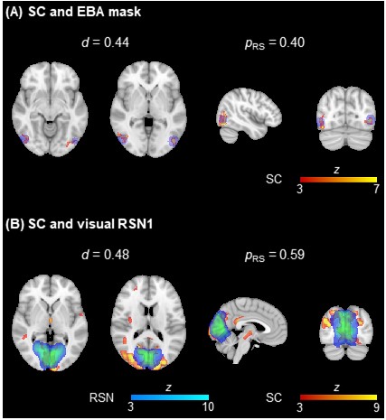
Fig. 3: Illustration of the overlap between two EEG SCs (in red-yellow) and (A)
the EBA mask (in blue) and (B) a visual RSN (in blue-light blue) from 15. The dice coefficient d and the proportion of the ROIs contained in the respective SCs
are also depicted.