Inês Esteves1, Ana R. Fouto1, Amparo Ruiz-Tagle1, Athanasios Vourvopoulos1, Marta Xavier1, Nuno A. Silva2, Raquel Gil-Gouveia3, Agostinho Rosa1, and Patrícia Figueiredo1
1ISR-Lisboa and Department of Bioengineering, Instituto Superior Técnico – Universidade de Lisboa, Lisbon, Portugal, 2Learning Health, Hospital da Luz, Lisbon, Portugal, 3Neurology Department, Hospital da Luz, Lisbon, Portugal
1ISR-Lisboa and Department of Bioengineering, Instituto Superior Técnico – Universidade de Lisboa, Lisbon, Portugal, 2Learning Health, Hospital da Luz, Lisbon, Portugal, 3Neurology Department, Hospital da Luz, Lisbon, Portugal
The feasibility of deriving ECG-derived respiration (EDR) signals for EEG-fMRI was shown, by comparison with the true respiratory signal. KPCA and PCA methods achieved the best performance, though EDR-based fMRI regressors should be further studied.
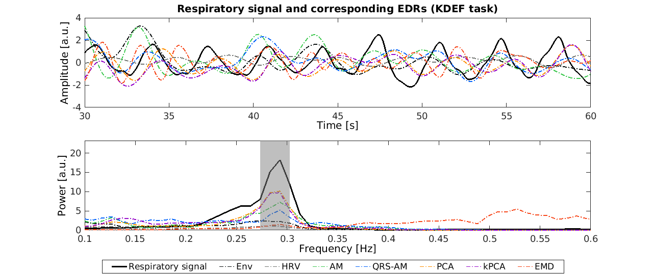
Figure 1: Respiratory signal and the corresponding EDRs obtained with different methods, from a representative subject performing the KDEF task: signal time courses over a period of 30s (top); and power spectra for the whole signal (bottom). The grey rectangle corresponds to the frequency band around the respiratory frequency for which the power is at least half of the maximum power.
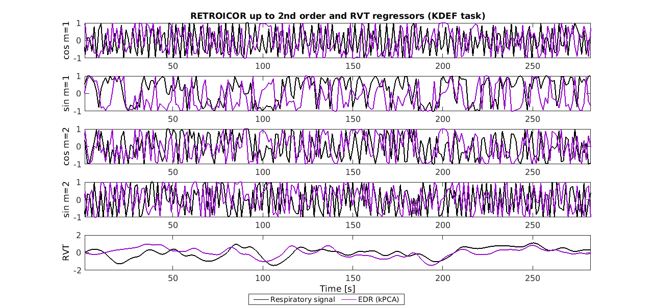
Figure 3: fMRI respiratory regressors, from a representative subject performing the KDEF task (same as in Fig.1): RETROICOR components obtained from the Fourier expansion up to the 2nd order of the respiratory phases (cosines and sines of order m=1,2); and respiratory volume per time (RVT), convolved with the respiratory response function [D], obtained from the measured respiratory signal (black) and the EDR signal estimated using the kPCA method (purple).