Hsin-Ju Lee1,2, Mikko Nyrhinen3, Risto J. Ilmoniemi3, and Fa-Hsuan Lin1,2,3
1Physical Sciences Platform, Sunnybrook Research Institute, Toronto, ON, Canada, 2Department of Medical Biophysics, University of Toronto, Toronto, ON, Canada, 3Department of Neuroscience and Biomedical Engineering, Aalto University, Espoo, Finland
1Physical Sciences Platform, Sunnybrook Research Institute, Toronto, ON, Canada, 2Department of Medical Biophysics, University of Toronto, Toronto, ON, Canada, 3Department of Neuroscience and Biomedical Engineering, Aalto University, Espoo, Finland
We measured fMRI signals caused by
excitatory and inhibitory TMS neuromodulations at the human primary motor
cortex. The primary motor cortex had fMRI signal after excitatory TMS. The
supplementary motor area had fMRI signals in both modulations.
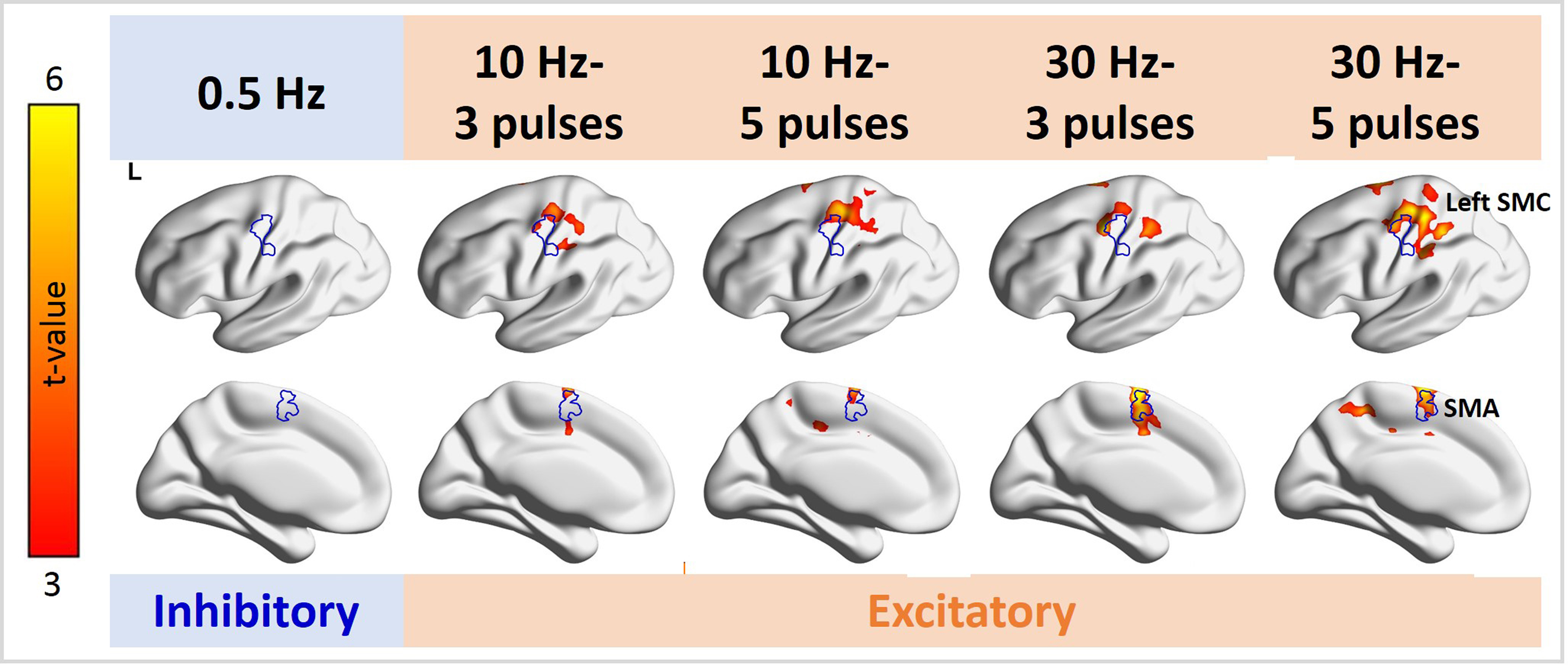
Figure 3. Distributions of significant fMRI signal changes in the individual TMS conditions. Positive fMRI signals were found at the SMC ipsilateral to the TMS target locus in all 10- and 30-Hz conditions, but not in the 0.5-Hz condition. The fMRI signal in the SMA was significantly increased in the 10-Hz–5-ppb, 30-Hz–3-ppb, and 30-Hz–5-ppb conditions.
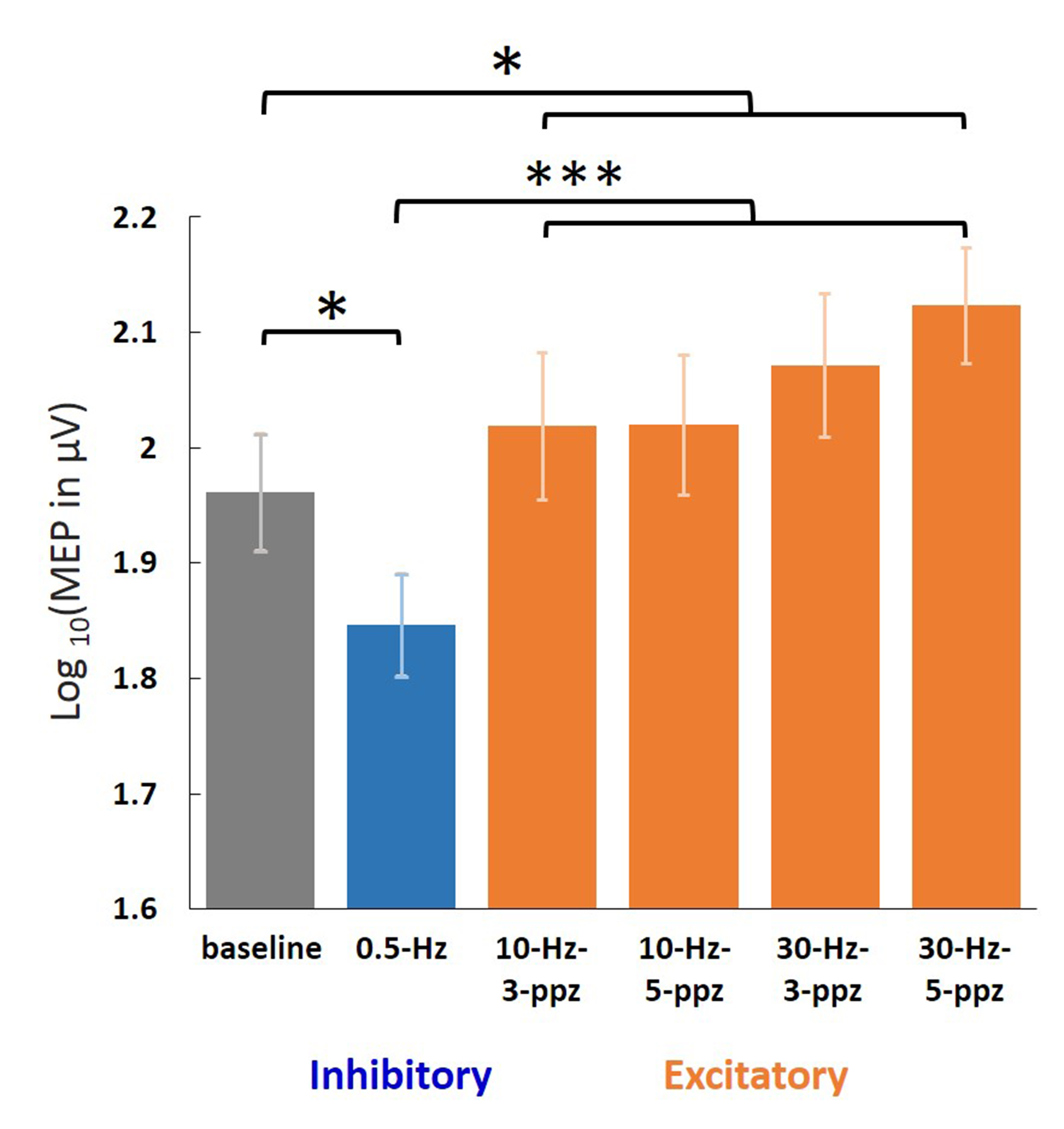
Figure 2. Log-transformed MEP amplitudes in the baseline
and in the individual TMS conditions. Significant MEPs were detected in all
conditions. The log-transformed MEP amplitude in the LF condition was
significantly smaller than in the baseline and HF conditions. In contrast, the
log-transformed MEP amplitude in the HF condition was significantly larger than
in the baseline. Error bars denote standard errors of the mean (SEM). *: p <
.05. ***: p < .001.