Thomas Lindner1, Hajrullah Ahmeti2, Michael Helle3, Olav Jansen4, Michael Synowitz2, and Stephan Ulmer4,5
1University Hospital Hamburg-Eppendorf, Hamburg, Germany, 2Neurosurgery, University Hospital Schleswig-Holstein, Kiel, Germany, 3Tomographic Imaging Department, Philips Research Laboratories, Hamburg, Germany, 4Department of Radiology and Neuroradiology, University Hospital Schleswig-Holstein, Kiel, Germany, 5Radiology, Kantonsspital Winterthur, Winterthur, Switzerland
1University Hospital Hamburg-Eppendorf, Hamburg, Germany, 2Neurosurgery, University Hospital Schleswig-Holstein, Kiel, Germany, 3Tomographic Imaging Department, Philips Research Laboratories, Hamburg, Germany, 4Department of Radiology and Neuroradiology, University Hospital Schleswig-Holstein, Kiel, Germany, 5Radiology, Kantonsspital Winterthur, Winterthur, Switzerland
During neurosurgery, it is crucial to spare functional areas. Thsi can be achieved by using perfusion iamging to visualize the tumor area as well as using resting state fMRI to map active regions. The presented technique allows for both in a single sequence.
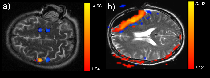
Figure 1: Representative examples of the activation of
the motor cortex (a) and false activation patterns in the resection cavity (b)
overlaid on T2 images.
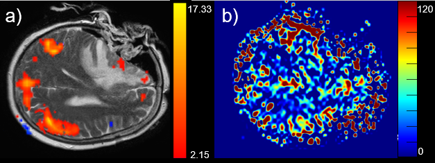
Figure 2: Example of the default mode network (a)
overlaid on a T2 image and the corresponding ASL perfusion (b). Note that in
the frontal area (medial prefrontal cortex and anterior cingulate cortex) no
default mode activation is visible as compared to the standard situation. The
inferior parietal cortex and cingulate cortex as well as the precuneus are visualized
as expected.