Kaden T Shearer1, Allen A Champagne2, Nicole S Coverdale1, Ingrid S Johnsrude3, and Douglas J Cook4
1Centre for Neuroscience Studies, Queen's University, Kingston, ON, Canada, 2Department of Medicine, Queen's University, Kingston, ON, Canada, 3The Brain and Mind Institute, University of Western Ontario, London, ON, Canada, 4Department of Surgery, Queen's University, Kingston, ON, Canada
1Centre for Neuroscience Studies, Queen's University, Kingston, ON, Canada, 2Department of Medicine, Queen's University, Kingston, ON, Canada, 3The Brain and Mind Institute, University of Western Ontario, London, ON, Canada, 4Department of Surgery, Queen's University, Kingston, ON, Canada
Compared to resting state, movie fMRI improves intersubject synchronization of the BOLD signal; however, no increases in BOLD-CBF coupling were observed. Improved ASL signal resolution and reduced noise may be required for differences in BOLD-CBF coupling to be revealed.
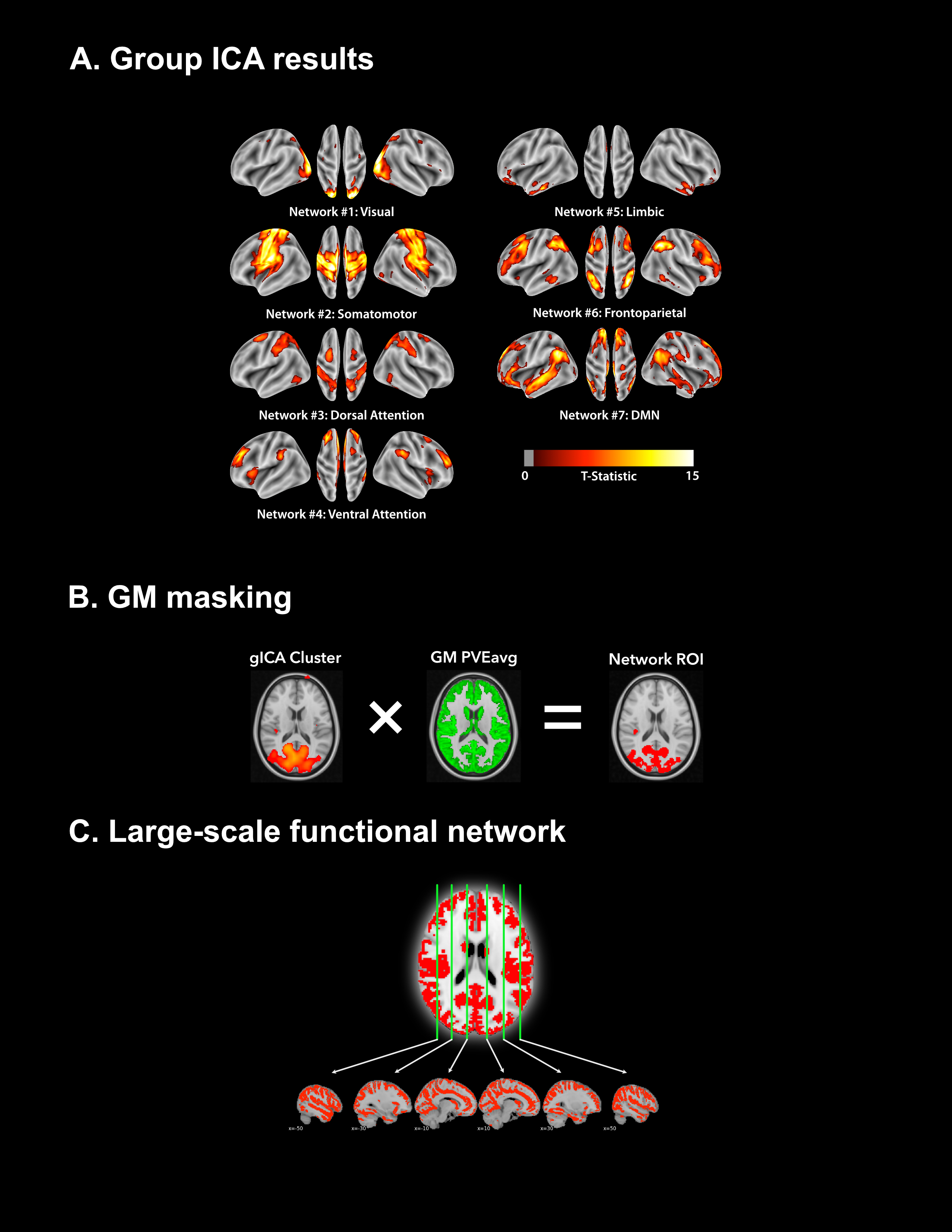
Figure 2. Schematic summarizing the workflow for establishing the large-scale functional ROI. (A) gICA was performed using FSL’s MELODIC11 with the number of pre-set spatial dimensions set to 50. Identified sub-networks were identified using the 7-network functional atlas13 and were thresholded at |t| > 4. (B) GM voxels were isolated by multiplying each identified sub-network by the subject’s GM partial volume estimation (PVE) mask, thresholded at 0.5. (C) The large-scale functional network mask was generated by spatially concatenating each identified GM sub-network.

Figure 4. Summary of intersubject BOLD synchronization for the various networks analyzed. In each correlation matrix, the rows/columns correspond to the average BOLD timeseries for a given subject. The values at each row/column intersection correspond to the Pearson R correlation between the BOLD signal for subject x and subject y. Correlation matrices were compared statistically using a univariate ANOVA (* = P < .05; ** = P < .01; *** = P < .001).