Mei-Yu Yeh1,2, Henry S Chen2, Ping Hou2, Vinodh A. Kumar3, Jason M Johnson3, Kyle R Noll4, Sujit S Prabhu5, Donald F. Schomer3, and Ho-Ling Liu 2
1Department of Biomedical Engineering and Environmental Sciences, National Tsing Hua University, Hsinchu, Taiwan, 2Department of Imaging Physics, The University of Texas MD Anderson Cancer Center, Houston, Houston, TX, United States, 3Departments of Diagnostic Radiology, The University of Texas MD Anderson Cancer Center, Houston, TX, United States, 4Department of Neuro-oncology, The University of Texas MD Anderson Cancer Center, Houston, TX, United States, 55Department of Neurosurgery, The University of Texas MD Anderson Cancer Center, Houston, TX, United States
1Department of Biomedical Engineering and Environmental Sciences, National Tsing Hua University, Hsinchu, Taiwan, 2Department of Imaging Physics, The University of Texas MD Anderson Cancer Center, Houston, Houston, TX, United States, 3Departments of Diagnostic Radiology, The University of Texas MD Anderson Cancer Center, Houston, TX, United States, 4Department of Neuro-oncology, The University of Texas MD Anderson Cancer Center, Houston, TX, United States, 55Department of Neurosurgery, The University of Texas MD Anderson Cancer Center, Houston, TX, United States
1.rRS (regression RS)-CVR showed better agreement with BH-CVR.2.The optimal frequent bands for rRS calculation in glioma patients are consistent with previous studies 3.The agreements between rRS- and BH-CVR were better in normal tissue than in the lesion.
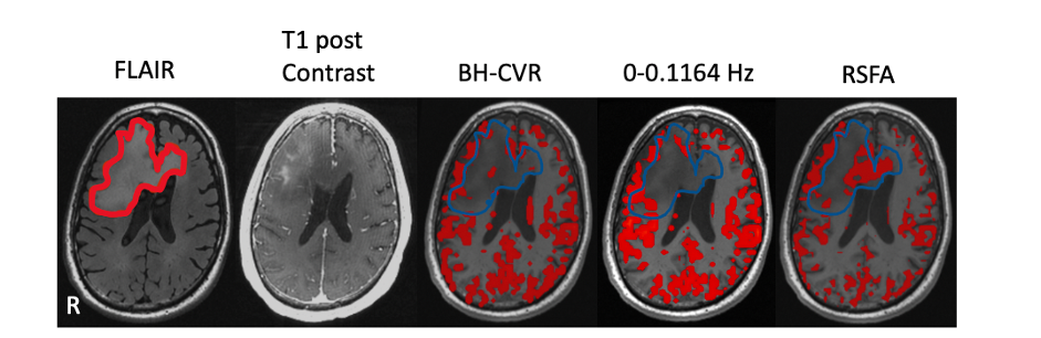
Fig.5 Imaging results from the one patient with gliomas. The first is lesion overlay on FLAIR image. The second one is T1 post contrast. The rest of all are spatial pattern among three CVR mapping approaches. The threshold of BH-CVR was set to 0.45. The threshold of rRS-CVR map was set to t>3.45(p<0.05). RSFA-CVR map was threshold such that the total activated fraction of gray matter in BH-CVR and RSFA map was equal.
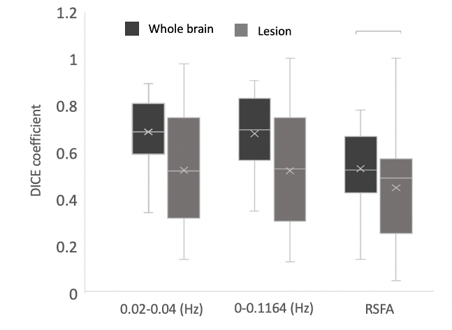
Fig.4 The dice coefficient between BH-CVR and three different methods of mapping CVR in normal brain tissue and in lesion (p<0.05).