Michael Bernier1,2, Jeorg Peter Pfannmoeller1,2, Saskia Bollmann3, Avery J.L. Berman1,2, and Jonathan R Polimeni1,2,4
1Department of Radiology, A. A. Martinos Center for Biomedical Imaging, Massachusetts General Hospital, Boston, MA, United States, 2Radiology, Harvard Medical School, Boston, MA, United States, 3Centre for Advanced Imaging, University of Queensland, Brisbane, Australia, 4Division of Health Sciences and Technology, Massachusetts Institute of Technology, Boston, MA, United States
1Department of Radiology, A. A. Martinos Center for Biomedical Imaging, Massachusetts General Hospital, Boston, MA, United States, 2Radiology, Harvard Medical School, Boston, MA, United States, 3Centre for Advanced Imaging, University of Queensland, Brisbane, Australia, 4Division of Health Sciences and Technology, Massachusetts Institute of Technology, Boston, MA, United States
We have developed
a “forward-model” method to calculate the extravascular fields surrounding the
blood vessels of the brain that accounts for the vessel diameter and
orientation and estimates the field change with activation using in vivo
measures of vessel anatomy and blood susceptibility.
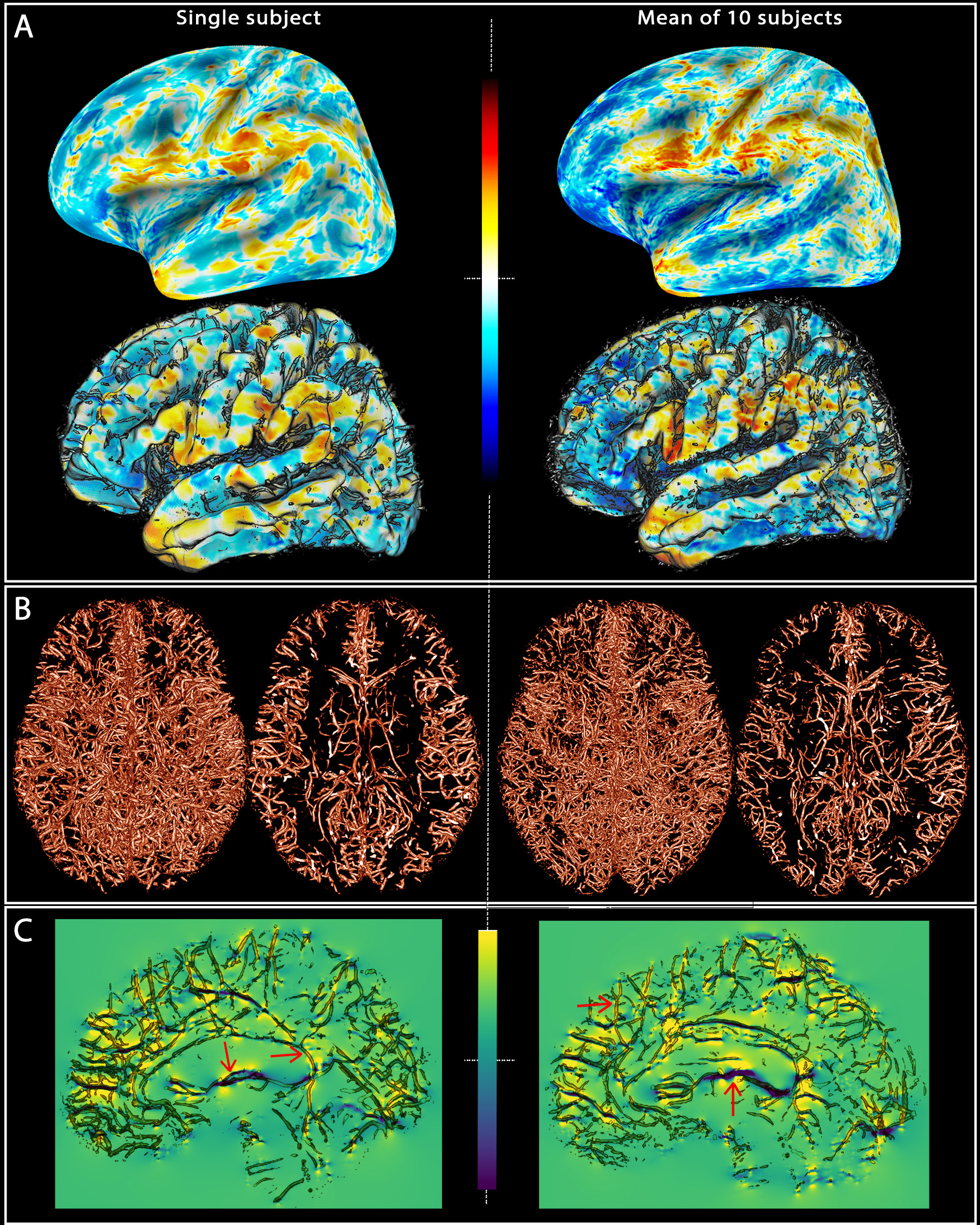
Fig. 1: Field offsets
surrounding major blood vessels The left panels illustrate the results for a single-subject while the right panels are the mean computed for all the subjects. (A) The field offset are projected on both inflated and GM surfaces obtained using Freesurfer. (B) The segmented vessels, illustrated in 3D and in a cross-section (20 mm), are overlapped on the delta B maps (C) to show the strong dipole effects surrounding the vessels perpendicular and parallel to B0 (red arrows).