Jacob Chausse1, Avery J. L. Berman2, and J. Jean Chen1,3
1Rotman Research Institute, Baycrest Health Sciences, North York, ON, Canada, 2A. A. Martinos Center for Biomedical Imaging, Harvard Medical School, Massachusetts General Hospital, Boston, MA, United States, 3Department of Medical Biophysics, University of Toronto, Toronto, ON, Canada
1Rotman Research Institute, Baycrest Health Sciences, North York, ON, Canada, 2A. A. Martinos Center for Biomedical Imaging, Harvard Medical School, Massachusetts General Hospital, Boston, MA, United States, 3Department of Medical Biophysics, University of Toronto, Toronto, ON, Canada
2D and 3D Monte Carlo simulations of the transverse relaxation rate show excellent agreement. This finding establishes the validity of the 2D approximation as a faster, less memory-intensive alternative.
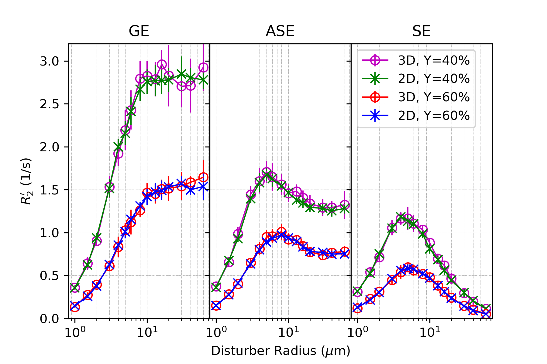
Figure 4. R2’ vs. Vessel Radius: comparing blood oxygenation with 2D MC and 3D MC for gradient echo (GE), asymmetric spin echo (ASE) and spin echo (SE). Uses CBV=2%, Hct=35.7%.
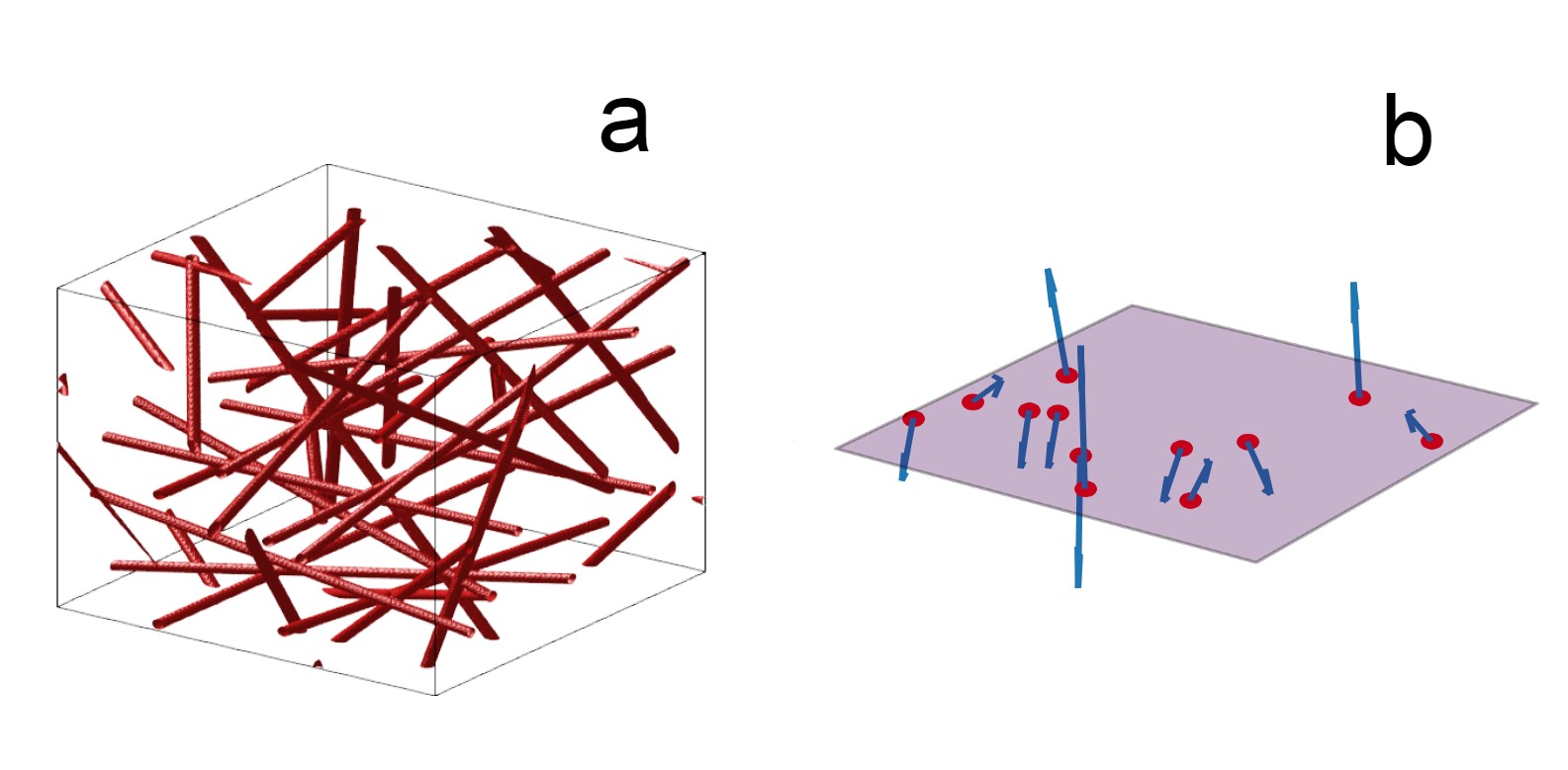
Figure 1. Visualization of the simulated voxels: (a) 3D voxel in a cube, where red cylinders represent blood vessels (b) 2D voxel on a plane where the vectors (blue) indicate the direction B0 at the vessel cross sections (red).