Khoi Minh Huynh1,2, Ye Wu2, and Pew-Thian Yap1,2
1Biomedical Engineering, UNC Chapel Hill, Chapel Hill, NC, United States, 2Department of Radiology and Biomedical Research Imaging Center (BRIC), UNC Chapel Hill, Chapel Hill, NC, United States
1Biomedical Engineering, UNC Chapel Hill, Chapel Hill, NC, United States, 2Department of Radiology and Biomedical Research Imaging Center (BRIC), UNC Chapel Hill, Chapel Hill, NC, United States
Microstructure fingerprinting improves estimation accuracy of axon/soma radii and membrane permeability without relying on simplifying assumptions associ- ated with conventional microstructure models.
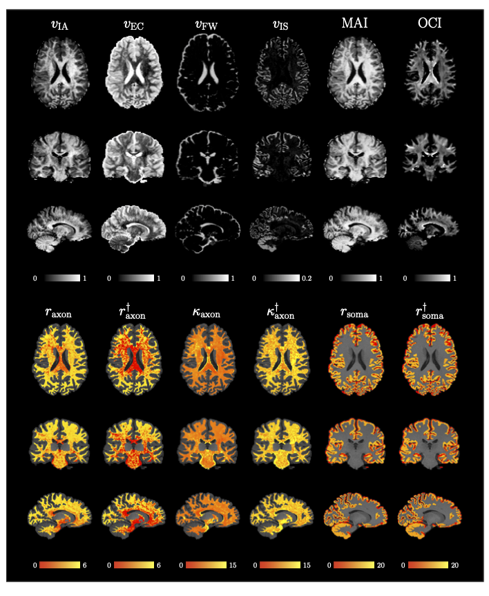
Fig. 4. MF-SMSI Indices. $$$v_{\text{IA}}$$$, $$$v_{\text{EC}}$$$, $$$v_{\text{FW}}$$$, $$$v_{\text{IS}}$$$, MAI, OCI, axonal radius $$$r_{\text{axon}}$$$ (μm), unbiased axonal radius $$$r_{\text{axon}}^\dagger$$$ (μm), axonaxonal permeability $$$\kappa_{\text{axon}}$$$ (10−6 μm μs−1), unbiased membrane permeability $$$\kappa_{\text{axon}}^\dagger$$$ (10−6 μm μs−1), soma radius $$$r_{\text{soma}}$$$ (μm), and unbiased soma radius $$$r_{\text{soma}}^\dagger$$$ (μm). Radius and permeability maps are overlaid on the T1w image.
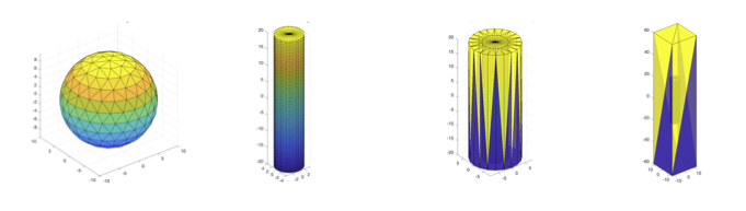
Fig. 1. Examples of SpinDoctor geometric configurations. From left to right: a sphere representing a soma with impermeable membrane a cylinder representing an axon with impermeable membrane, a cylinder tightly wrapped with extra-cellular space, and a cylinder in a boxed extra-cellular space representing an axon with perme- able membrane allowing water exchange with extra-cellular space.