Steven H. Baete1,2, Patryk Filipiak1,2, Lee Basler3, Anthony Zuccolotto3, Ying-Chia Lin1,2, Dimitris G. Placantonakis4, Timothy Shepherd1,2, Walter Schneider3, and Fernando E. Boada1,2
1Center for Advanced Imaging Innovation and Research (CAI2R), NYU School of Medicine, New York, NY, United States, 2Center for Biomedical Imaging, Dept. of Radiology, NYU School of Medicine, New York, NY, United States, 3Psychology Software Tools, Inc., Pittsburgh, PA, United States, 4Department of Neurosurgery, Perlmutter Cancer Center, Neuroscience Institute, Kimmel Center for Stem Cell Biology, NYU School of Medicine, New York, NY, United States
1Center for Advanced Imaging Innovation and Research (CAI2R), NYU School of Medicine, New York, NY, United States, 2Center for Biomedical Imaging, Dept. of Radiology, NYU School of Medicine, New York, NY, United States, 3Psychology Software Tools, Inc., Pittsburgh, PA, United States, 4Department of Neurosurgery, Perlmutter Cancer Center, Neuroscience Institute, Kimmel Center for Stem Cell Biology, NYU School of Medicine, New York, NY, United States
Orientation
Distribution Functions derived from several methods were directly compared
using a ground truth phantom imaged with clinical 3T MRI. Results demonstrate difficulties shared across conventional methods
resolving fiber crossing angles less than 45°.
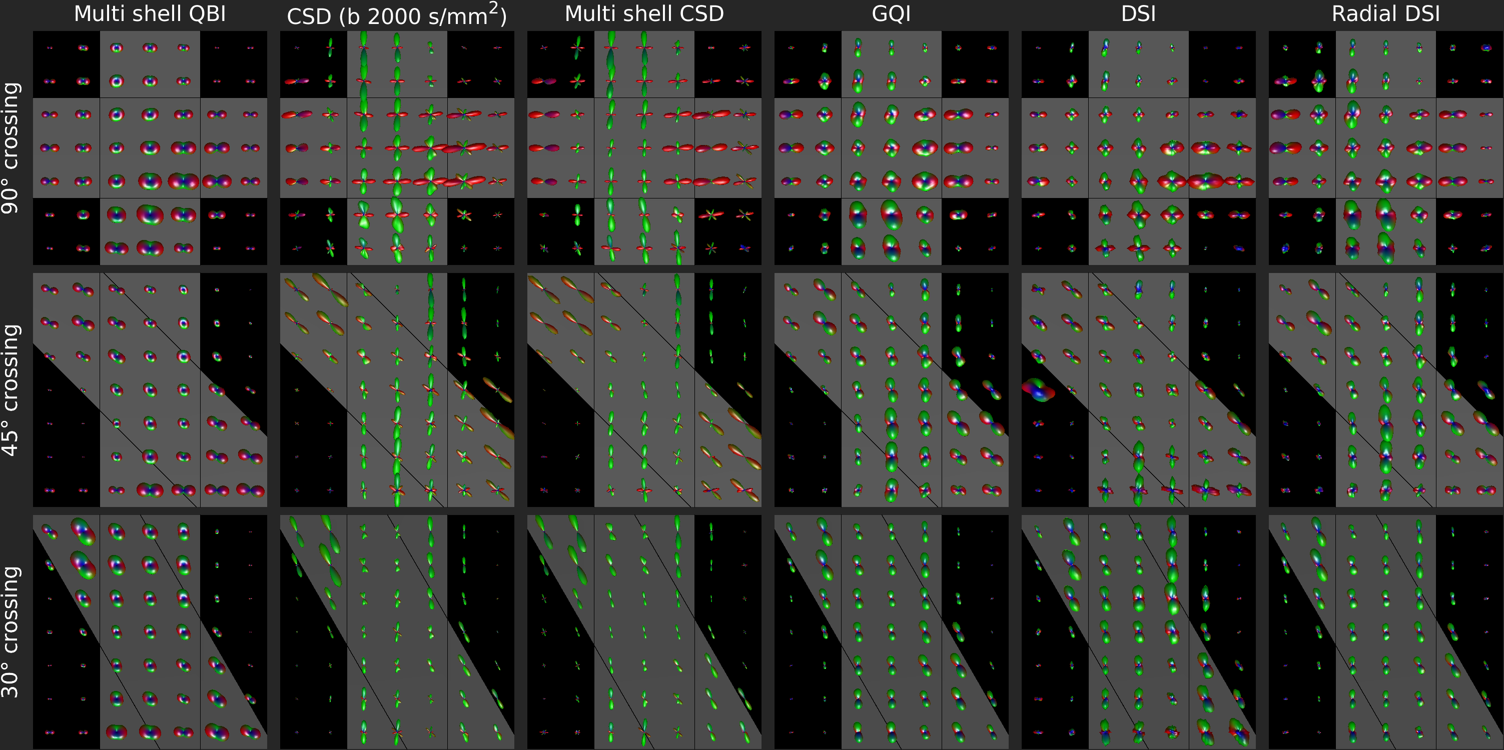
Figure 3:
ODFs in ROIs of Taxon bundles crossing at 90°, 45° and 30° (rows) as
reconstructed by Multi-shell QBI (qODF), CSD (fODF), GQI (dODF), DSI (dODF) and
RDSI (dODF) (columns). Gray bands represent ground truth fiber directions.
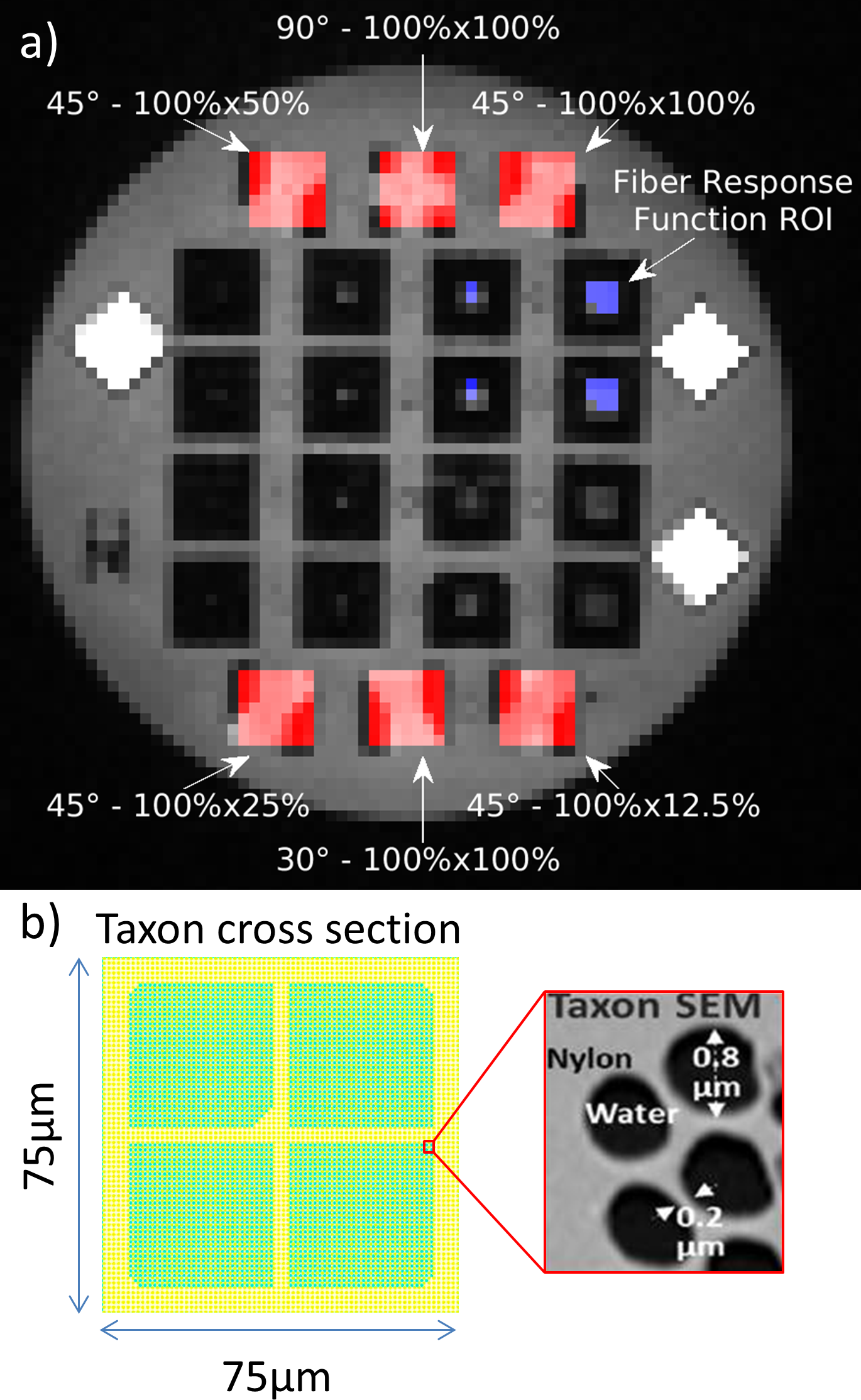
Figure 1: The anisotropic
diffusion phantom contains single and crossing bundles of Taxons™ (textile
water filled tubes [9] with 0.8 10-6 microtubes). a) T1-weighted image of the phantom layout. The ROIs used
in this work are indicated along with crossing fiber angles and taxon
densities. b) Taxon cross section schematic and SEM of Taxon detail.