Arthur Chakwizira1, Filip Szczepankiewicz2, Linda Knutsson1,3, Pia Sundgren2,4,5,6, and Markus Nilsson2
1Department of Medical Radiation Physics, Lund University, Lund, Sweden, 2Department of Diagnostic Radiology, Lund University, Lund, Sweden, 3Russell H. Morgan Department of Radiology and Radiological Science, Johns Hopkins University School of Medicine, Baltimore, MD, United States, 4Lund University Bioimaging Center, Lund University, Lund, Sweden, 5Department for Medical Imaging and Physiology, Skåne University Hospital, Lund, Sweden, 6Department of Radiology, University of Michigan, Ann Arbor, MI, United States
1Department of Medical Radiation Physics, Lund University, Lund, Sweden, 2Department of Diagnostic Radiology, Lund University, Lund, Sweden, 3Russell H. Morgan Department of Radiology and Radiological Science, Johns Hopkins University School of Medicine, Baltimore, MD, United States, 4Lund University Bioimaging Center, Lund University, Lund, Sweden, 5Department for Medical Imaging and Physiology, Skåne University Hospital, Lund, Sweden, 6Department of Radiology, University of Michigan, Ann Arbor, MI, United States
We demonstrated that our previously proposed unified framework is applicable for measuring restricted diffusion and exchange in vivo. Pilot experiments in a healthy brain and a meningioma patient indicate plausible parameter estimates.
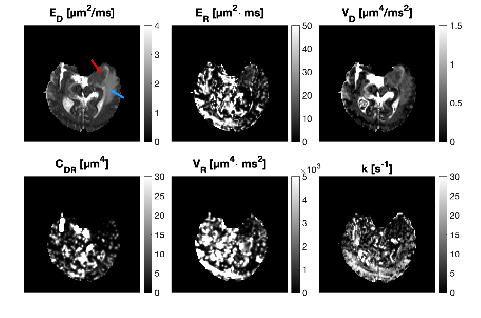
Figure 4: Parametric maps resulting from fitting equation 1 on data obtained on a meningioma patient. The tumour (located in the left temporal lobe) is marked with a red arrow and the oedema with a blue arrow. $$$E_D, E_R, V_D, C_{D,R}$$$ and $$$V_R$$$ denote mean diffusivity, mean restriction coefficient, variance of diffusivities, covariance of $$$D$$$ and $$$R$$$ and the variance of $$$R$$$, respectively. In addition to noise, there is a pronounced fat shift artefact at the bottom of the maps.
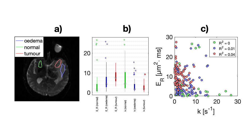
Figure 5: (a): Slice through the brain of a meningioma patient showing three ROIs placed in normal tissue and the tumour as well as the oedema around it. (b): Variation of the restriction coefficient and exchange rate in the three regions. The former increases in the tumour relative to normal tissue, while the latter decreases. (c): Quantification of the correlation between restriction and exchange in the three regions. The plot shows lack of correlation, meaning the parameters convey independent information.