Hong Jiang1, João Pedro de Almeida Martins1, Dan Lundberg2, Chantal M. W. Tax3, and Daniel Topgaard1
1Physical Chemistry, Lund University, Lund, Sweden, 2CR Competence AB, Lund, Sweden, 3Cardiff University Brain Research Imaging Centre (CUBRIC), Cardiff University, Cardiff, United Kingdom
1Physical Chemistry, Lund University, Lund, Sweden, 2CR Competence AB, Lund, Sweden, 3Cardiff University Brain Research Imaging Centre (CUBRIC), Cardiff University, Cardiff, United Kingdom
This paper presents a lamellar liquid crystal phantom for use on clinical scanners for validation of advanced diffusion encoding methods to disambiguate truly planar from "crossing fibers" tissue microstructures that give similar signal response on conventional DTI.
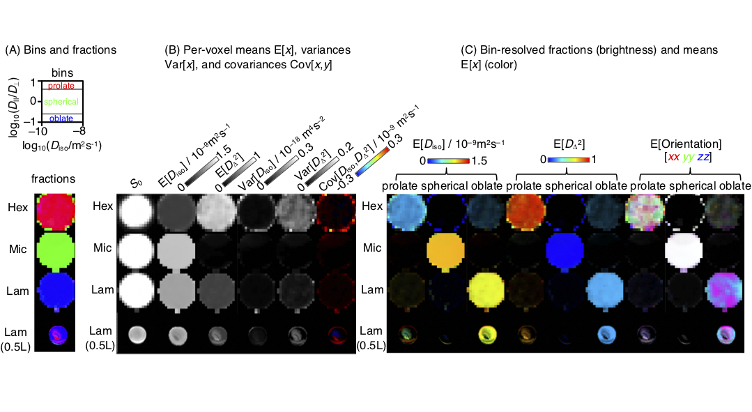
Figure 2. Parameter maps derived from the per-voxel D-distributions for the 5 mm Hex, Mic, and Lam phantoms for microimaging and the 0.5 L Lam phantom on the whole-body scanner. (A) Image segmentation by binning in the 2D Diso-DA/DR projection to capture tensor components characteristic of Hex (prolate), Mic (spherical), and Lam (oblate). (B) Per-voxel statistical measures E[x], Var[x] and Cov[x,y] of the Diso and DΔ2 projections of the distributions. (C) Bin-resolved signal fractions (brightness) and per-bin means (color). Ideal lamellar gives rise to foblate = 1 and DΔ2 = 0.25.