Elisabetta Pagani1, Paolo Preziosa1,2, Raffaello Bonacchi1,2, Laura Cacciaguerra1,2,3, Massimo Filippi1,2,3,4,5, and Maria A. Rocca1,2,3
1Neuroimaging Research Unit, Division of Neuroscience, IRCCS San Raffaele Scientific Institute, Milan, Italy, 2Neurology Unit, IRCCS San Raffaele Scientific Institute, Milan, Italy, 3Vita-Salute San Raffaele Unversity, Milan, Italy, 4Neurorehabilitation Unit, IRCCS San Raffaele Scientific Institute, Milan, Italy, 5Neurophysiology Service, IRCCS San Raffaele Scientific Institute, Milan, Italy
1Neuroimaging Research Unit, Division of Neuroscience, IRCCS San Raffaele Scientific Institute, Milan, Italy, 2Neurology Unit, IRCCS San Raffaele Scientific Institute, Milan, Italy, 3Vita-Salute San Raffaele Unversity, Milan, Italy, 4Neurorehabilitation Unit, IRCCS San Raffaele Scientific Institute, Milan, Italy, 5Neurophysiology Service, IRCCS San Raffaele Scientific Institute, Milan, Italy
A significant neurite loss occurs in the cortex
of multiple sclerosis patients, being more severe with longer disease duration
and more severe disability. Cortical lesions show a further reduction, with
increased inflammation, gliosis, and
simplification of neurite complexity.
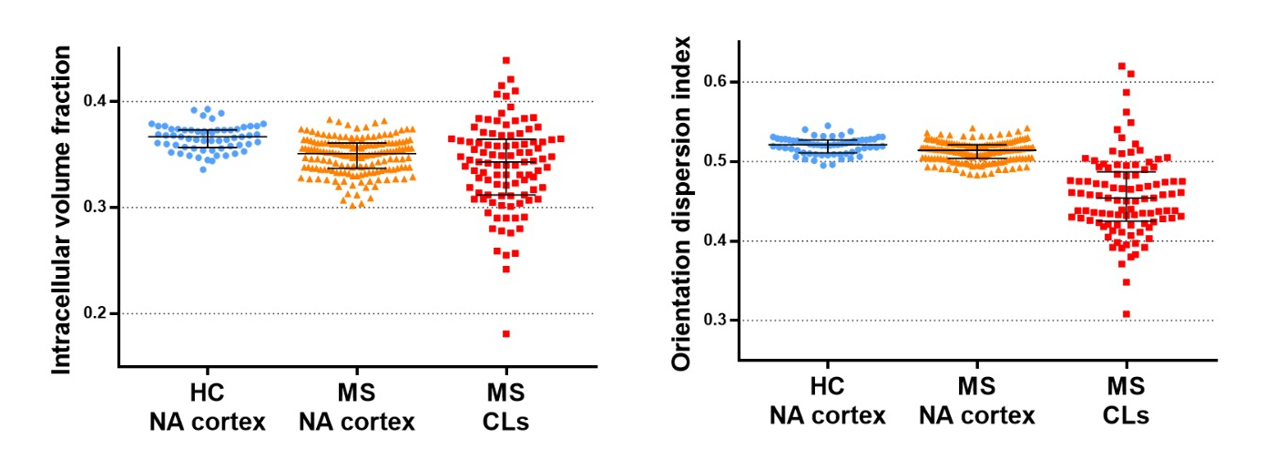
Figure 2. Scatter plots of NODDI
indexes obtained within the normal-appearing cortex (NA-cortex) and cortical
lesions (CLs) of MS patients and healthy controls.
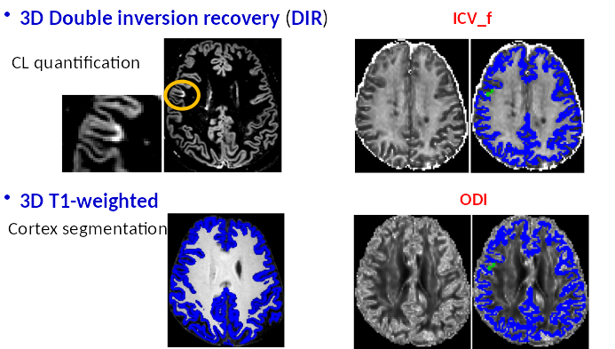
Figure 1. The postprocessing is
shown for a representative MS patient: after the segmentation of cortical
lesions and gray matter, masks are overlapped on the maps of intracellular
volume fraction (ICV_f) and orientation dispersion (ODI).