Susanne Rauh1, Oliver Maier2, Oliver Gurney-Champion3, Melissa Hooijmans3, Rudolf Stollberger2,4, Aart Nederveen3, and Gustav Strijkers1
1Department of Biomedical Engineering and Physics, Amsterdam UMC, location AMC, Amsterdam, Netherlands, 2Institute of Medical Engineering, Graz University of Technology, Graz, Austria, 3Department of Radiology and Nuclear Medicine, Amsterdam UMC, location AMC, Amsterdam, Netherlands, 4BioTechMed-Graz, Graz, Austria
1Department of Biomedical Engineering and Physics, Amsterdam UMC, location AMC, Amsterdam, Netherlands, 2Institute of Medical Engineering, Graz University of Technology, Graz, Austria, 3Department of Radiology and Nuclear Medicine, Amsterdam UMC, location AMC, Amsterdam, Netherlands, 4BioTechMed-Graz, Graz, Austria
Model-based reconstruction is feasible for IVIM and combined
IVIM-DTI fitting in abdominal organs. The parameter maps reveal more detail and show less artifacts compared to those obtained with a conventional fit. Mean values are similar between the two methods.
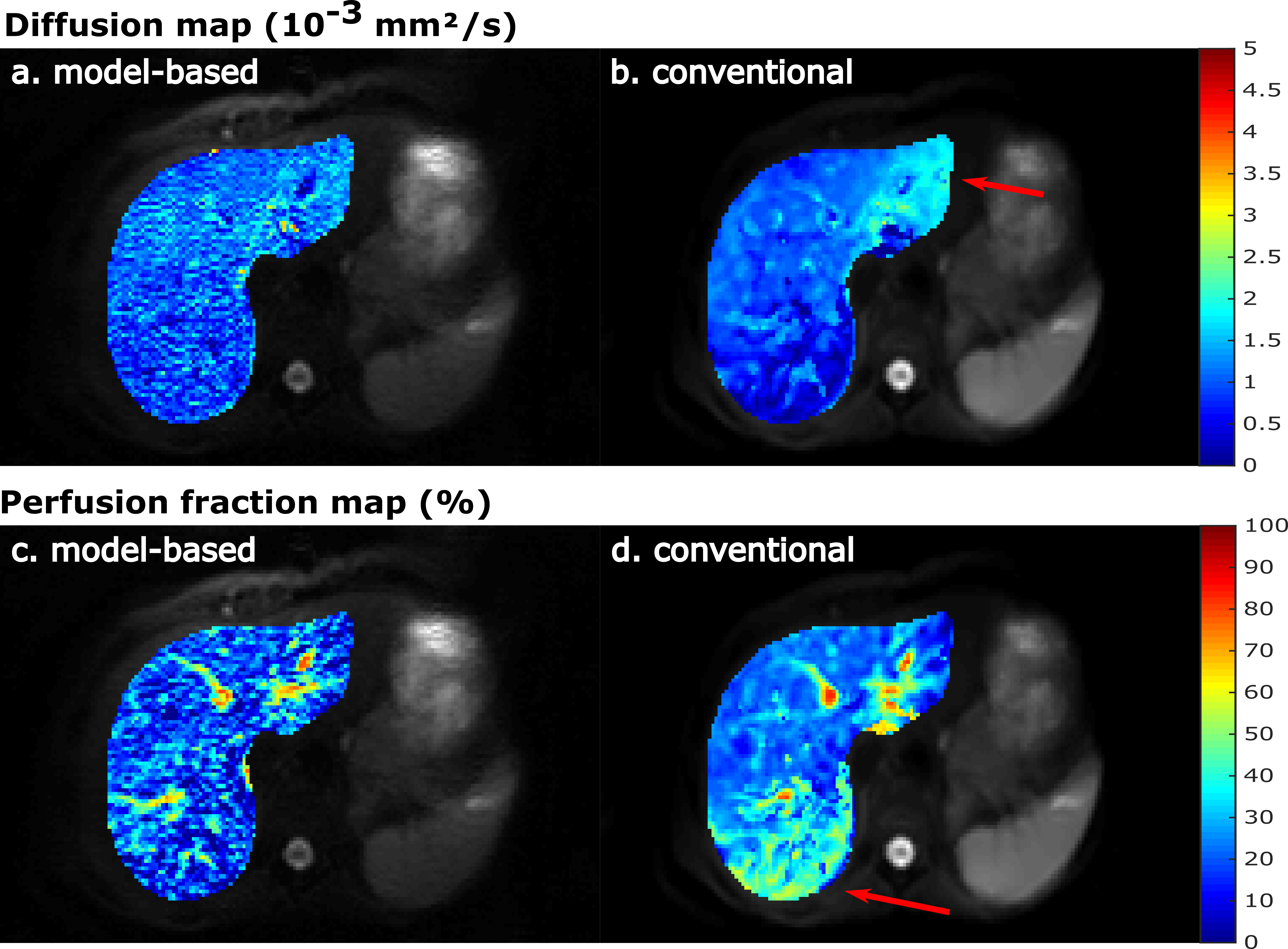
Figure 3: IVIM
parameter maps of the liver, obtained with model-based reconstruction on the
left (a and c) and the conventional fit on the right-hand side (b and d). The
conventional fit seems to overestimate the diffusion coefficient in the left
liver lobe and perfusion fraction in the bottom of the liver (red arrows). This
behavior is not observed in the model-based reconstructed maps.
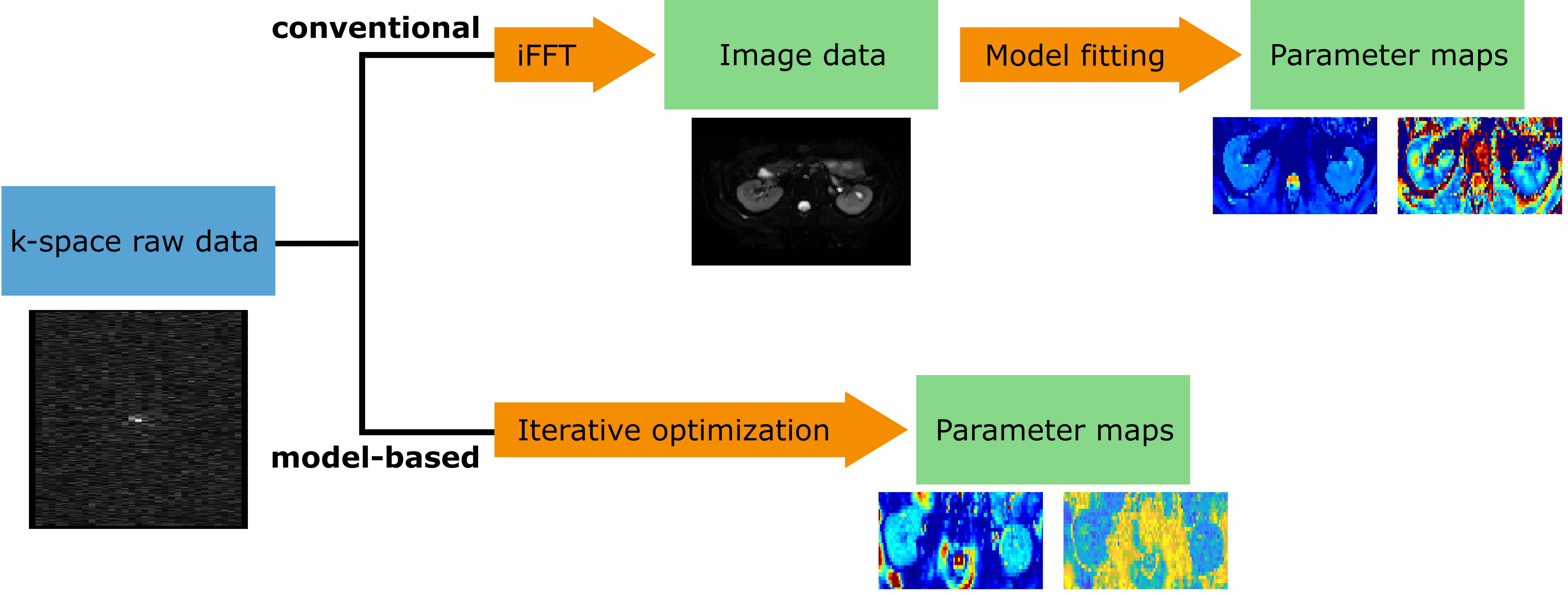
Figure 1: Schematic view of the conventional (top) and
model-based (bottom) reconstruction and fitting process. Conventionally,
magnitude-only images are reconstructed prior to the model fitting. However,
the modulus operation transforms the noise from a complex Gaussian to a Rician
distribution. In the model-based reconstruction the quantitative model is
included in the reconstruction process, thus the Gaussian noise assumption is
valid. iFFT: inverse fast Fourier transform.