Jens T Rosenberg1, Samuel Colles Grant1,2, and Daniel Topgaard3
1National High Magnetic Field Laboratory, Florida State University, Tallahassee, FL, United States, 2Chemical and Biomedical Engineering, FAMU-FSU College of Engineering, Tallahassee, FL, United States, 3Physical Chemistry, Lund University, Lund, Sweden
1National High Magnetic Field Laboratory, Florida State University, Tallahassee, FL, United States, 2Chemical and Biomedical Engineering, FAMU-FSU College of Engineering, Tallahassee, FL, United States, 3Physical Chemistry, Lund University, Lund, Sweden
Here we implement multidimensional diffusion-relaxation correlation methods at 21.1 T. Results are reproducible compared to lower field strengths but with reduced image quality due to increase in R2, showing the need for stronger gradient to shorten the duration of
gradient waveforms
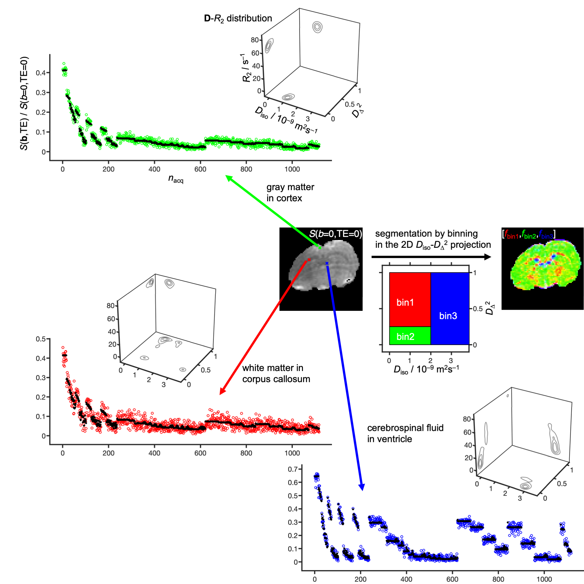
Signal S vs.
acquisition number nacq (circles: measured, points: fit) and
nonparametric D-R2 distributions for representative
voxels of an in vivo rat brain at 21.1 T. The distributions are
shown as projections onto the 2D planes Diso-DΔ2, Diso-R2,
and DΔ2-R2,
where Diso is the isotropic diffusivity, DΔ2 the squared normalized anisotropy (41), and R2 the transverse
relaxation rate. Binning in the Diso-DΔ2 plane allows calculation of nominally
tissue-specific signal fractions fbin1, fbin2,
and fbin3 and associated diffusion-relaxation metrics.
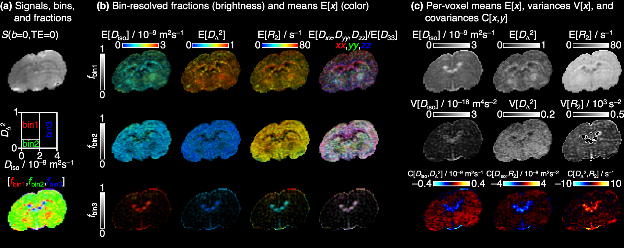
Parameter maps derived from the per-voxel D-R2
distributions. (a) Synthesized S(b=0,TE=0) image, bin
definition in the Diso-DΔ2 plane, and RGB map color-coded by the signal
fractions [fbin1,fbin2,fbin3].
(b) Bin-resolved signal fractions and means E[x] of the D-R2
metrics coded into image brightness and color (see quantitative scale bars). Direction-encoded
colors derive from the lab-frame diagonal values [Dxx,Dyy,Dzz]
and maximum eigenvalue D33. (c) Per-voxel means E[x],
variances V[x], and covariances C[x,y] of Diso,
DΔ2, and R2.