Vanessa Wiggermann1, Henrik Lundell1, Mads Alexander Just Madsen1, Christopher Fugl Madelung1, and Hartwig Roman Siebner1,2,3
1Danish Research Centre for Magnetic Resonance, Centre for Functional and Diagnostic Imaging and Research, Copenhagen University Hospital Hvidovre, Hvidovre, Denmark, 2Dept. of Neurology, Copenhagen University Hospital Bispebjerg, Copenhagen, Denmark, 3Institute for Clinical Medicine, Faculty of Medical and Health Sciences, University of Copenhagen, Copenhagen, Denmark
1Danish Research Centre for Magnetic Resonance, Centre for Functional and Diagnostic Imaging and Research, Copenhagen University Hospital Hvidovre, Hvidovre, Denmark, 2Dept. of Neurology, Copenhagen University Hospital Bispebjerg, Copenhagen, Denmark, 3Institute for Clinical Medicine, Faculty of Medical and Health Sciences, University of Copenhagen, Copenhagen, Denmark
We showed that automated spinal cord measurements in healthy controls, multiple sclerosis and Parkinson’s patients at 7T are consistent between segmentation tools and with 3T data, if images with similar voxel sizes are used. At higher spatial resolution, smaller area estimates are obtained.
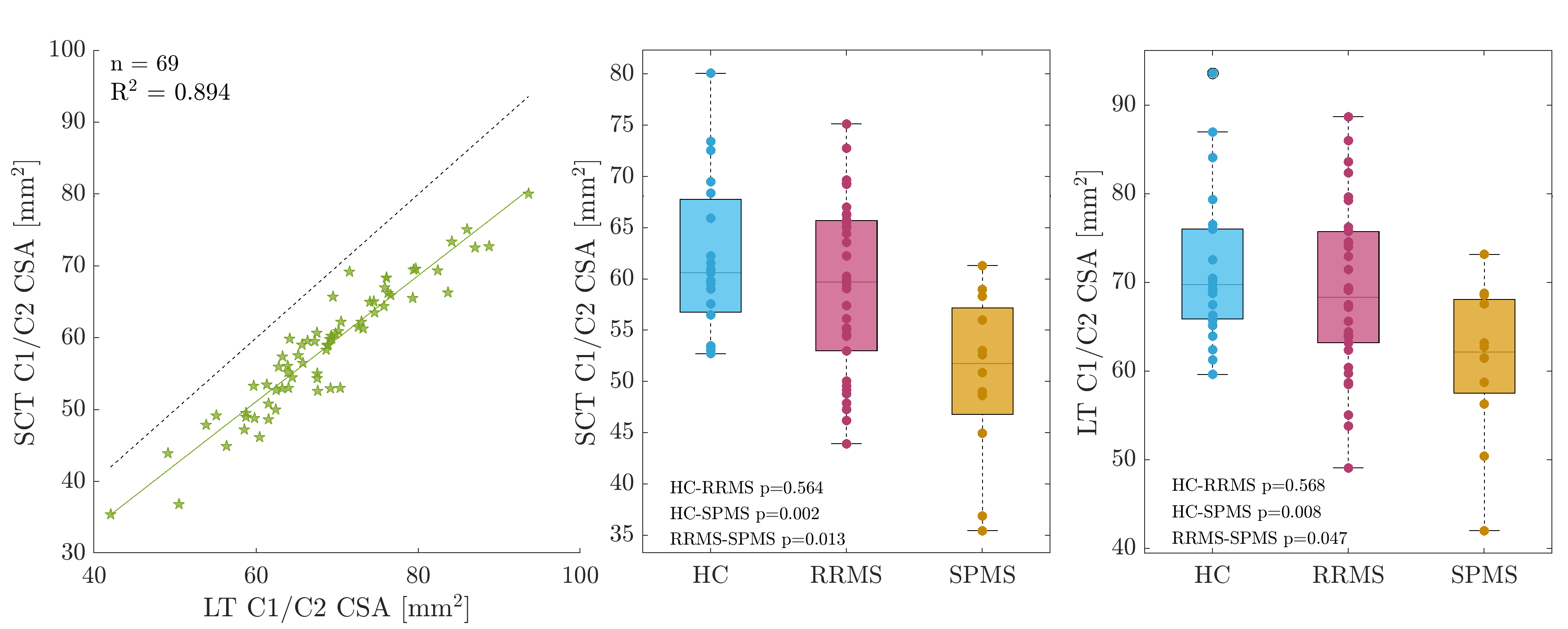
Figure 2: Comparison of average CSA measurements at the C1/C2 level using SCT and the in-house technique (LT). Although SCT generally obtained lower CSA measurements, the relative reproducibility of CSA was very high, both on an individual level (see left regression of individual data) and on a group level (compare middle and right). Statistical analysis involved a non-parametric Kruskal-Wallis test with post-hoc multiple comparison correction by Tukey-Kramer. P-values are uncorrected for testing both SCT and LT.
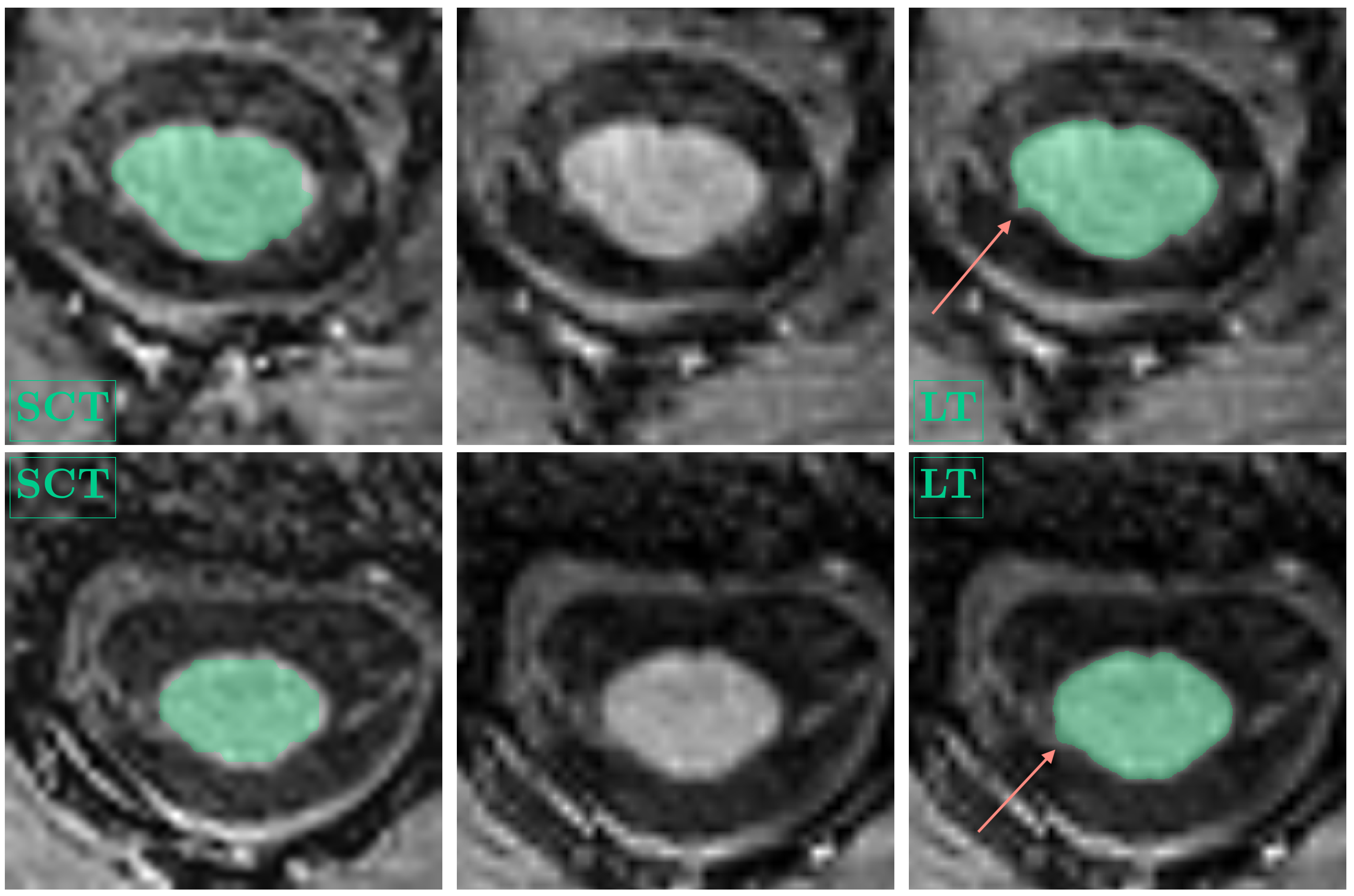
Figure 1: Example spinal cord segmentations at the C1/C2 level, SCT (left), LT (right). MPRAGE images without overlaid segmentation are shown for reference in the center. For display purposes, the SCT segmentations were upsampled to the LT standard and slices were visually matched. Arrows point toward partial segmentation of spinal nerve roots. For subject 1, (top row, female, RRMS, 33 years), mean SCT-CSA was 65.04 mm2, compared to LT-CSA=74.03 mm2. For subject 2, (bottom row, female, HC, 42 years), mean SCT-CSA was 53.49 mm2, compared to LT-CSA=61.26 mm2.