1Biophysics, Medical College of Wisconsin, Milwaukee, WI, United States, 2Neurosurgery, Medical College of Wisconsin, Milwaukee, WI, United States
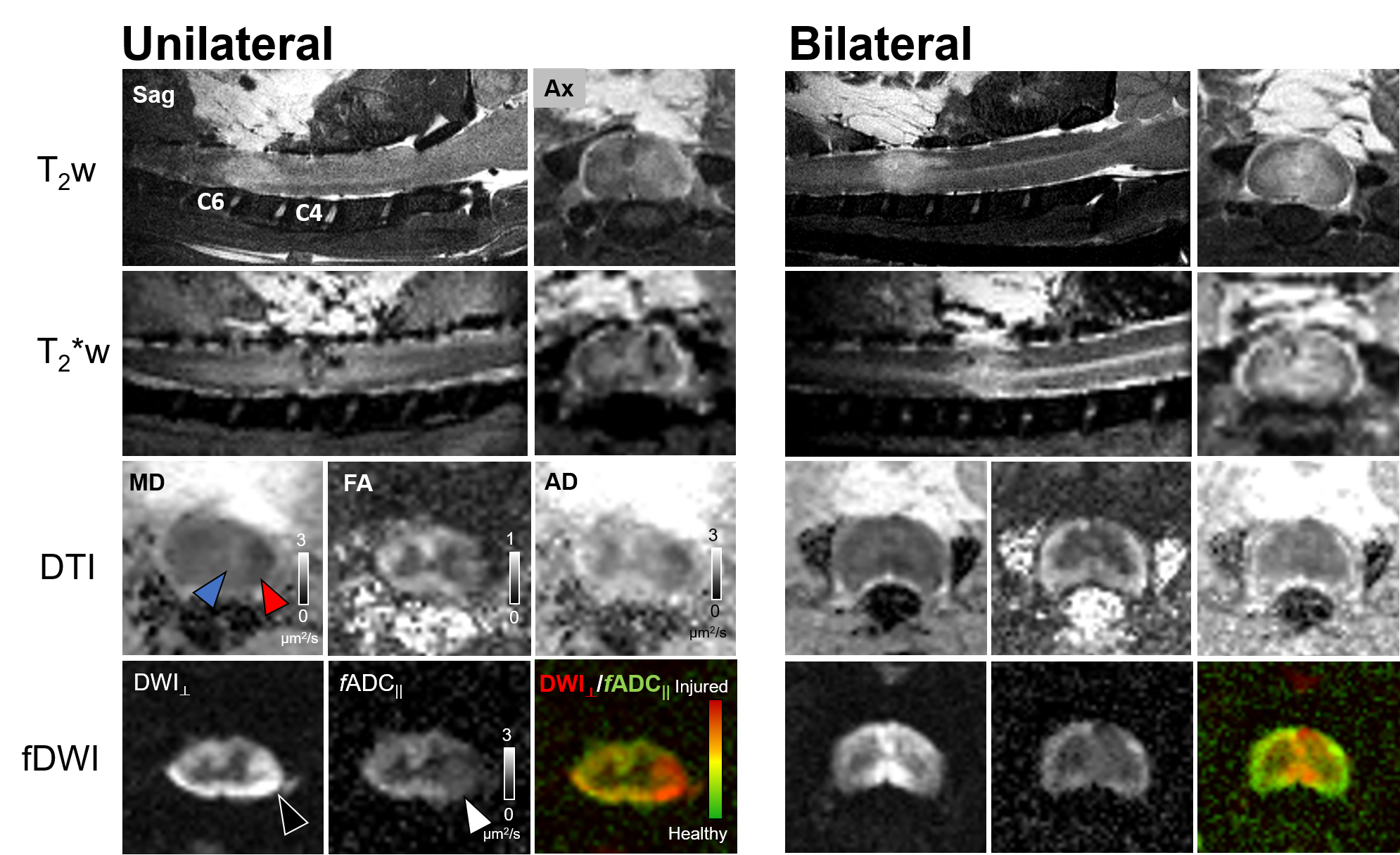
Figure 1 Multi-contrast MRI in acute cervical cord contusion
The extent of the cord damage is captured in different modalities in both unilateral- and bilateral contusion injury. Edema (hyperintensity, T2w), hemorrhage (hypointensity, T2*w), vasogenic edema (blue, black triangle), and cytotoxic edema (red, white triangle) are evident in their respective contrasts. fDWI images clearly delineates focal axonal damage compared to T2w, T2*w and DTI.
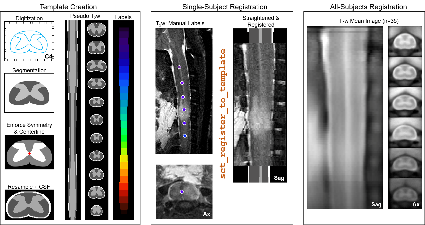
Figure 2 Inter-subject registration pipeline
A histologic spinal cord atlas was digitized across all spinal cord levels to generate a template with pseudo T2-weighted contrast used for MRI registration. Manually marked spinal cord level aided initial z-level alignment and straightening followed by x-y plane registration to the template. Registered single subject images were averaged and registered iteratively across all subjects with the group mean image showing clear spinal cord anatomy.
