Anna JE Combes1,2, Kristin P O'Grady1,2, Baxter P Rogers1,2, Kurt G Schilling1,2, Richard D Lawless2,3, Mereze Visagie2, Delaney Houston2, Colin D McKnight1, Francesca R Bagnato4, John C Gore1,2,3,4, and Seth A Smith1,2,3
1Radiology & Radiological Sciences, Vanderbilt University Medical Center, Nashville, TN, United States, 2Vanderbilt University Institute of Imaging Science, Vanderbilt University Medical Center, Nashville, TN, United States, 3Biomedical Engineering, Vanderbilt University, Nashville, TN, United States, 4Neurology, Vanderbilt University Medical Center, Nashville, TN, United States
1Radiology & Radiological Sciences, Vanderbilt University Medical Center, Nashville, TN, United States, 2Vanderbilt University Institute of Imaging Science, Vanderbilt University Medical Center, Nashville, TN, United States, 3Biomedical Engineering, Vanderbilt University, Nashville, TN, United States, 4Neurology, Vanderbilt University Medical Center, Nashville, TN, United States
DTI and resting-state fMRI were acquired in the cervical cord in a relapsing-remitting MS group with low disability. Higher functional connectivity (FC) in the dorsal network was correlated with DTI markers of tissue damage, suggesting increased FC may be a pathological feature in this group.
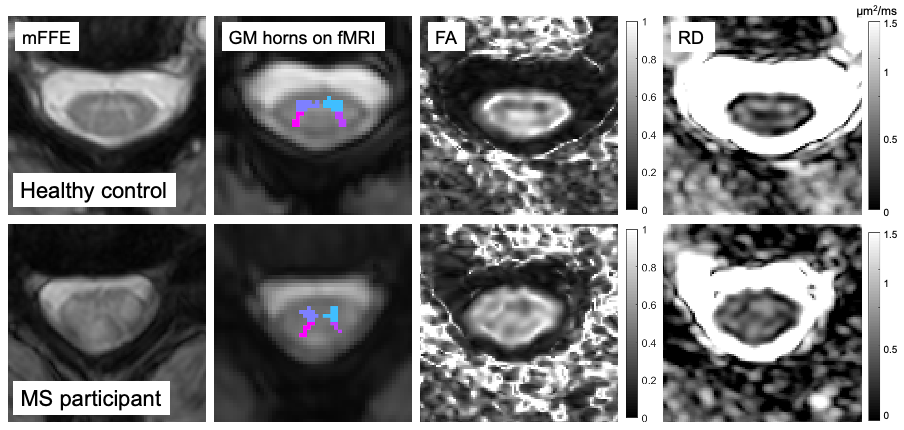
Figure 2. Example slices for a control (M, 35 years old) and a participant with MS (F, 42 years old, EDSS 1.5). Left to right: anatomical T2*-weighted mFFE for segmentation; grey matter horns regions of interest overlaid on average functional run; FA and RD diffusion maps. FA = Fractional Anisotropy. GM = Grey Matter. RD = Radial Diffusivity.
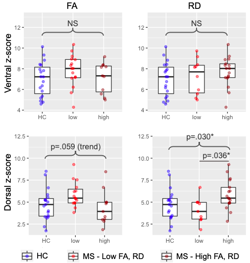
Figure 4. Ventral and dorsal network z-scores by subgroups: healthy controls (HC), MS with low FA/RD, MS with high FA/RD. FA = Fractional Anisotropy. RD = Radial Diffusivity. NS = Non Significant. *Significant at p<.05.