Arash Forodighasemabadi1,2,3,4, Henitsoa Rasoanandrianina1,2,3,4, Mohamed Mounir El Mendili1,2, Maxime Guye1,2, and Virginie Callot1,2,4
1Aix-Marseille Univ, CNRS, CRMBM, Marseille, France, 2APHM, Hopital Universitaire Timone, CEMEREM, Marseille, France, 3Aix-Marseille Univ, Université Gustave Eiffel, LBA, Marseille, France, 4iLab-Spine International Associated Laboratory, Montreal, Canada, Marseille, France
1Aix-Marseille Univ, CNRS, CRMBM, Marseille, France, 2APHM, Hopital Universitaire Timone, CEMEREM, Marseille, France, 3Aix-Marseille Univ, Université Gustave Eiffel, LBA, Marseille, France, 4iLab-Spine International Associated Laboratory, Montreal, Canada, Marseille, France
This work proposes an optimized MP2RAGE protocol and postprocessing pipeline for simultaneous brain and cervical spinal cord (BCSC) T1 mapping at 3T, providing large spatial coverage, high CNR, low B1+ sensitivity, and short acquisition time, while benefiting from high reproducibility.
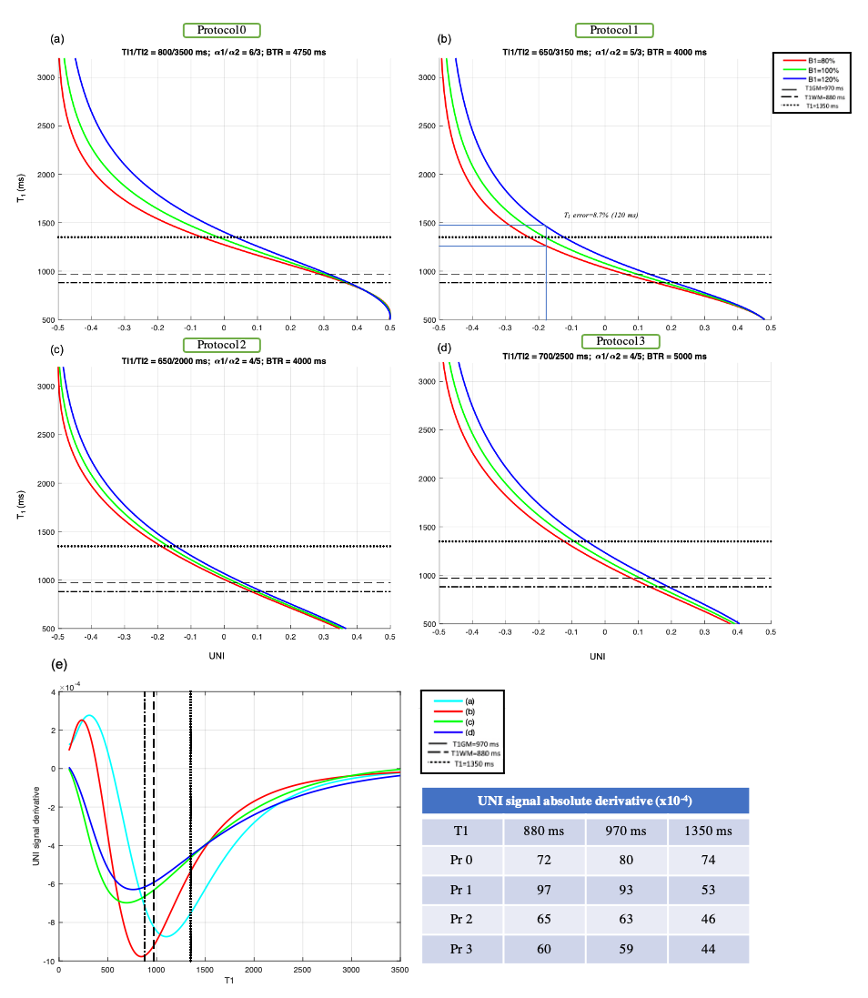
Figure 1: Relationships between UNI signal and estimated T1 value. (a) The protocol Pr0, provides the highest CNR at the expense of acquisition time. (b) The protocol Pr1 provides a high CNR, while allowing to cover brain and CSC in less than 8 minutes. It should be noted that a ±20% B1+ variation for a T1 of 1350 (brain GM) for instance, leads to an estimation error of 8.7%, showing the necessity for B1+ correction. Protocols previously optimized for (c) studying brain2,14, and (d) SC7, independently. (e) Derivative of the UNI signal, providing an indication on tissue discrimination.
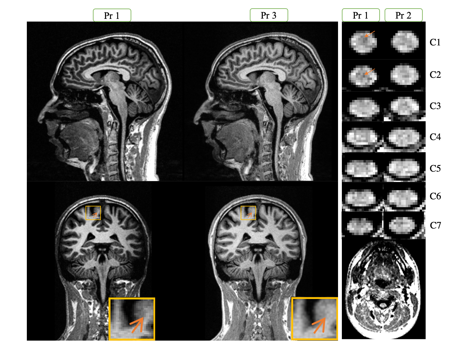
Figure 3 : A representation of the UNI-denoised image (no unit) for Pr1 and 3 on brain and Pr1 and 2 along the cervical SC. In the bottom-right image we can see a zoomed-out section of spine, which shows the need for sub-millimetric resolution for imaging SC. We can observe a nice delineation between different structures of brain and SC, with a higher signal difference (contrast) between GM and WM for the optimized protocol 1 (arrows).