Bragi Sveinsson1,2, Robert L Barry1,2,3, Olivia Rowe1,2, Jason Stockmann1,2, Daniel J Park1, Peter J Lally4, Matthew S Rosen1,2,5, and Reza Sadjadi6
1Athinoula A. Martinos Center for Biomedical Imaging, Massachusetts General Hospital, Boston, MA, United States, 2Radiology, Harvard Medical School, Boston, MA, United States, 3Harvard-Massachusetts Institute of Technology Health Sciences and Technology, Cambridge, MA, United States, 4Brain Sciences, Imperial College London, London, United Kingdom, 5Physics, Harvard University, Cambridge, MA, United States, 6Neurology, Massachusetts General Hospital, Boston, MA, United States
1Athinoula A. Martinos Center for Biomedical Imaging, Massachusetts General Hospital, Boston, MA, United States, 2Radiology, Harvard Medical School, Boston, MA, United States, 3Harvard-Massachusetts Institute of Technology Health Sciences and Technology, Cambridge, MA, United States, 4Brain Sciences, Imperial College London, London, United Kingdom, 5Physics, Harvard University, Cambridge, MA, United States, 6Neurology, Massachusetts General Hospital, Boston, MA, United States
Quantitative Double-Echo in Steady-State imaging at 7 Tesla with a 28-channel QED coil enables sub-150 μm anatomical and diffusion-weighted imaging of individual peripheral nerve fascicles.
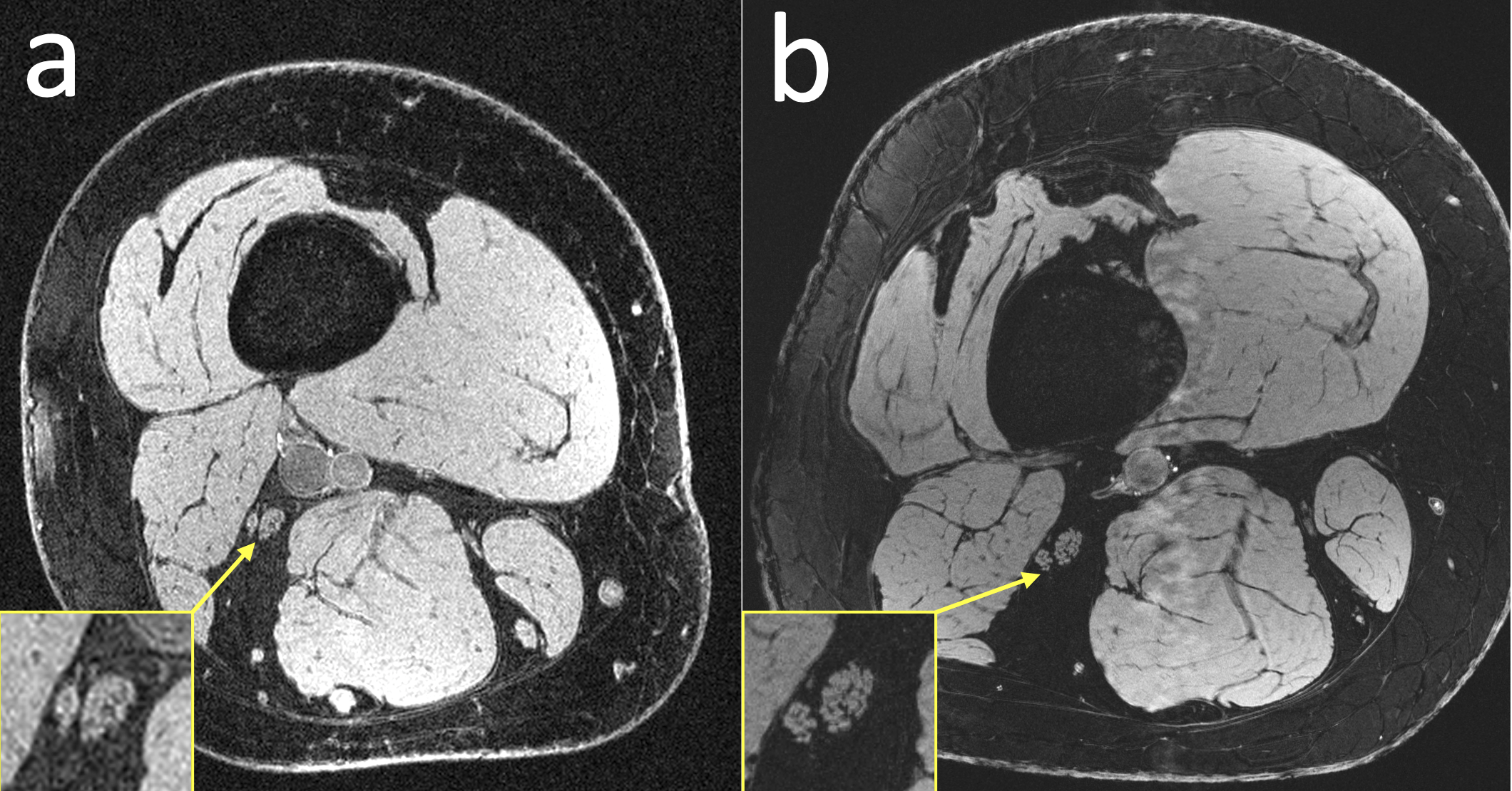
Figure 3: Anatomical comparison of a healthy subject scanned (a) at 3T with a 4-channel coil that wrapped around the thigh and (b) at 7T with a 28-channel QED coil. The individual fascicles of the sciatic nerves are clearly visible in the 7T image, but are more difficult to make out in the 3T image.
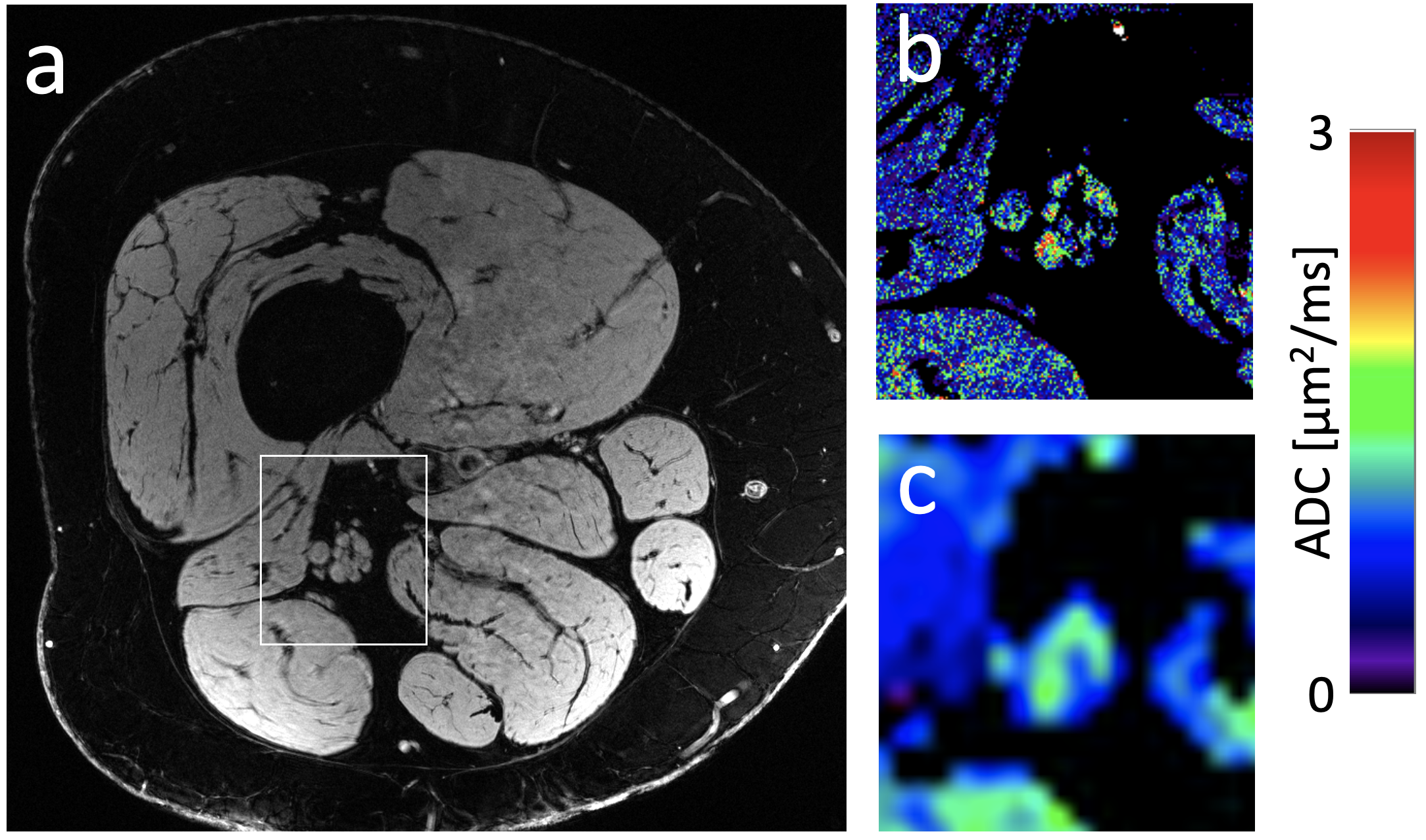
Figure 4: (a) A 7T image of a subject with peripheral neuropathy and (b) a DESS ADC map computed in the framed section of panel a. (c) A conventional EPI DWI diffusion map for comparison. The EPI map not only has worse resolution but also has severe distortion.