Kurt G. Schilling1,2, Shreyas Fadnavis3, Mereze Visagie2, Eleftherios Garyfallidis3, Bennett A. Landman2,4, Seth A. Smith1,2, and Kristin P. O'Grady1,2
1Radiology and Radiological Sciences, Vanderbilt University Medical Center, Nashville, TN, United States, 2Vanderbilt University Institute of Imaging Science, Vanderbilt University Medical Center, Nashville, TN, United States, 3Intelligent Systems Engineering, Indiana University Bloomington, Bloomington, IN, United States, 4Electrical Engineering and Computer Science, Vanderbilt University, Nashville, TN, United States
1Radiology and Radiological Sciences, Vanderbilt University Medical Center, Nashville, TN, United States, 2Vanderbilt University Institute of Imaging Science, Vanderbilt University Medical Center, Nashville, TN, United States, 3Intelligent Systems Engineering, Indiana University Bloomington, Bloomington, IN, United States, 4Electrical Engineering and Computer Science, Vanderbilt University, Nashville, TN, United States
We used Patch2Self to denoise spinal cord diffusion MRI data which is inherently limited in SNR with few gradient directions. Using self-supervised machine learning, we improved inter- and intra-session repeatability and conspicuity of pathology.
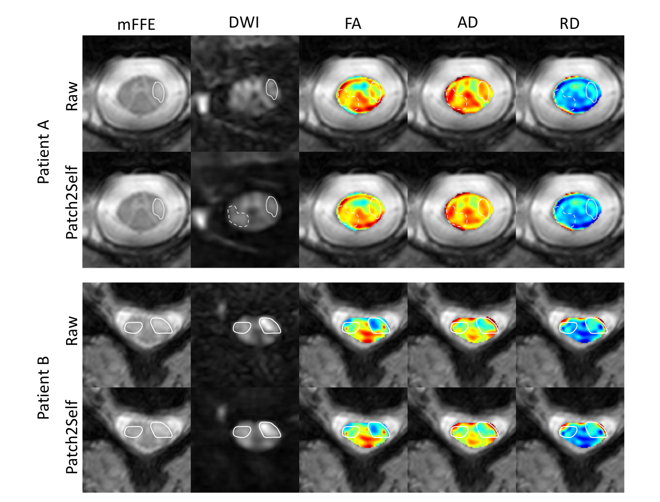
Figure 5: Two representative MS patients: Patient A (25yr female, EDSS=0) and Patient B (38yr female, EDSS=1) without (top) and with (bottom) Patch2Self denoising applied to the dMRI data. Lesions in the lateral columns (white outlines) of both patients are more conspicuous in the denoised diffusion volumes, including one lesioned area (dashed outline) that is not apparent in the corresponding mFFE anatomical image for Patient A.
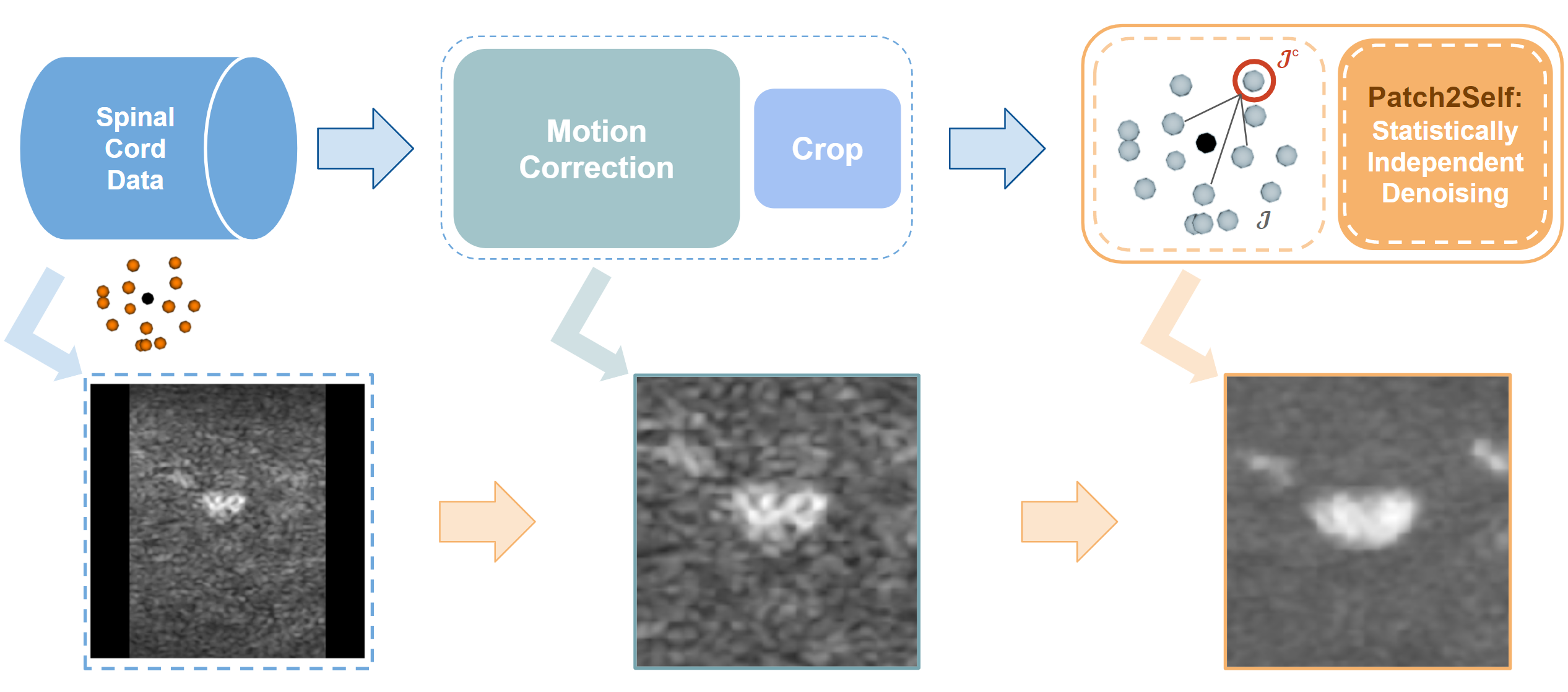
Figure 1: Flow diagram of pre-processing steps for Patch2Self denoising of spinal cord diffusion MRI data.