Kevin M Koch1 and Andrew S Nencka1
1Radiology, Medical College of Wisconsin, Milwaukee, WI, United States
1Radiology, Medical College of Wisconsin, Milwaukee, WI, United States
Technical methods are described and demonstrated that allow for automated advanced morphological analysis of the spinal cord in the presence of stabilization instrumentation.
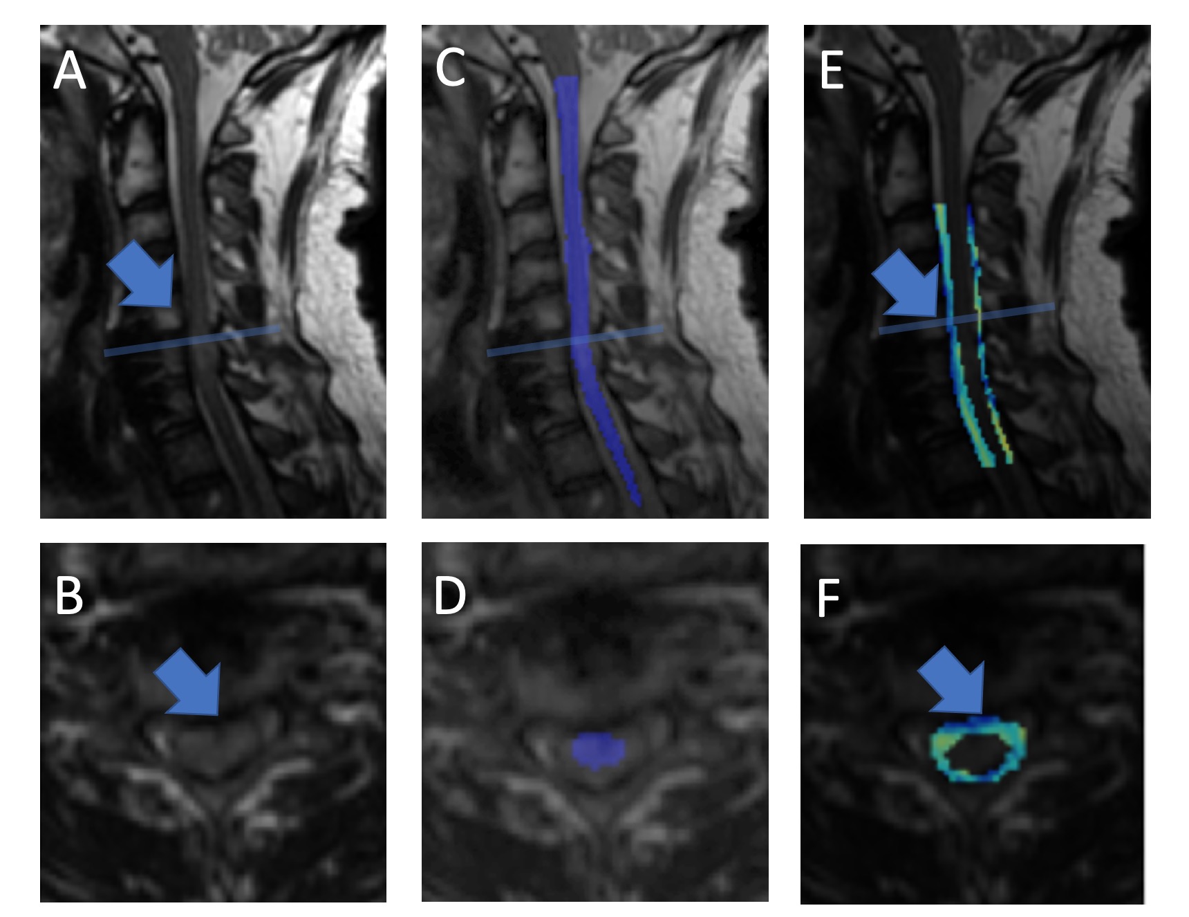
Figure 4. Example results of the described pipeline. A) and B) provide reformats of the T2w 3D-MSI, while C-D) provide views of the automatically segmented cord performed using the workflow outlined in Figure 3. E-F) provide maps of CSF z-score analysis, whereby the mean T2w signal of the CSF (corrected for signal shading an bias within the cord) is used to identify regions of low CSF (blue areas in the map). The blue arrows highlight a region of missing CSF (cord impingement), which is accurately reflected by the sharp blue region in the z-score map.
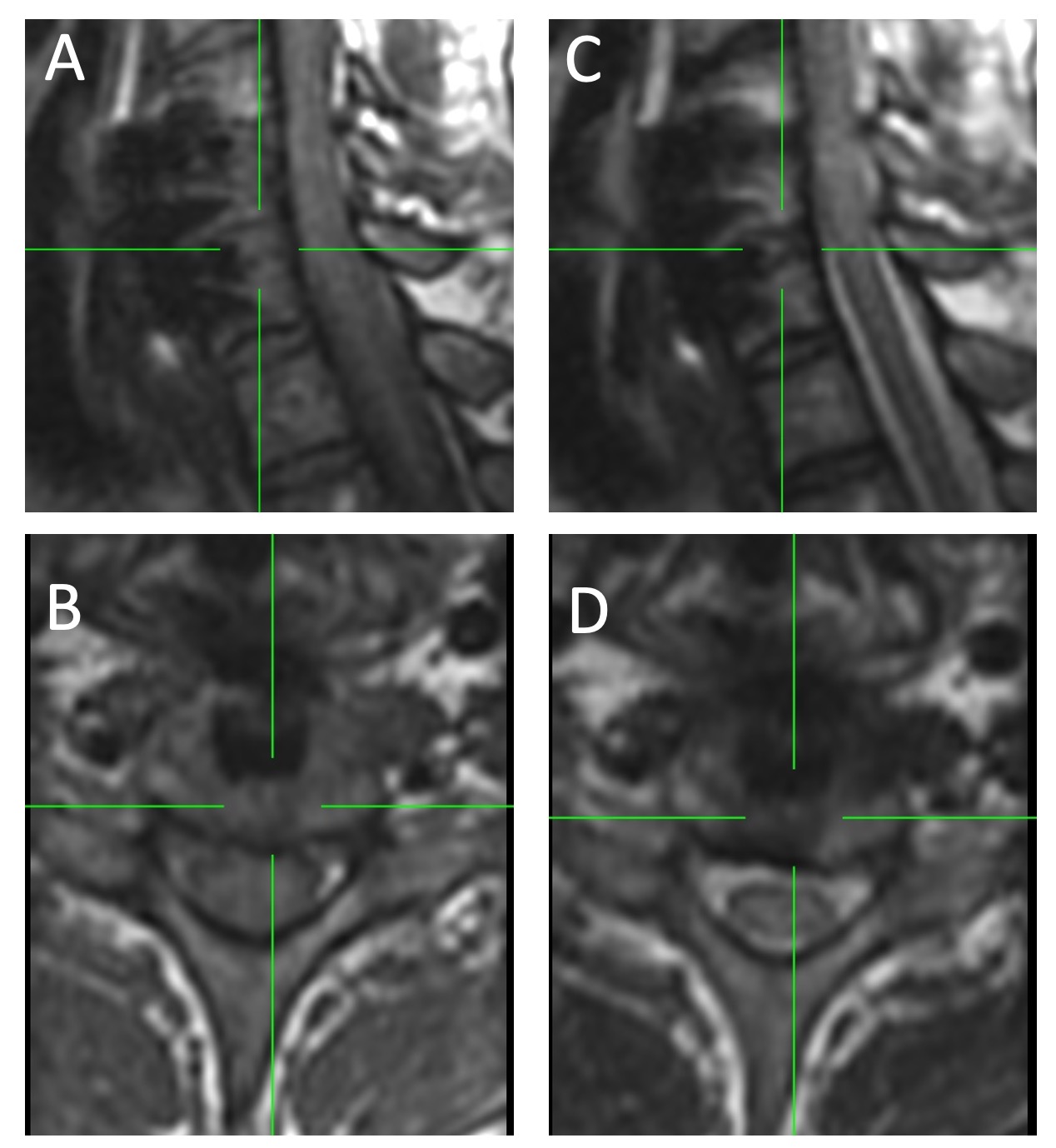
Figure 2: Example T1 (A,B) and T2 (C,D) weighted 1.2 mm isotropic acquisitions in orthogonal reformatted (sagittal, A,C – axial, B,D) view planes. The isotropic acquisitions required 5 minutes each and were designed for morphological cord analysis rather than conventional planar diagnostics.