Jian Wang1, Yanjun Chen1, Yingjie Mei2, Jialing Chen1, and Xiaodong Zhang1
1Department of Medical Imaging, The Third Affiliated Hospital of Southern Medical University, Guangzhou, China, 2China International Center, Philips Healthcare, Guangzhou, China
1Department of Medical Imaging, The Third Affiliated Hospital of Southern Medical University, Guangzhou, China, 2China International Center, Philips Healthcare, Guangzhou, China
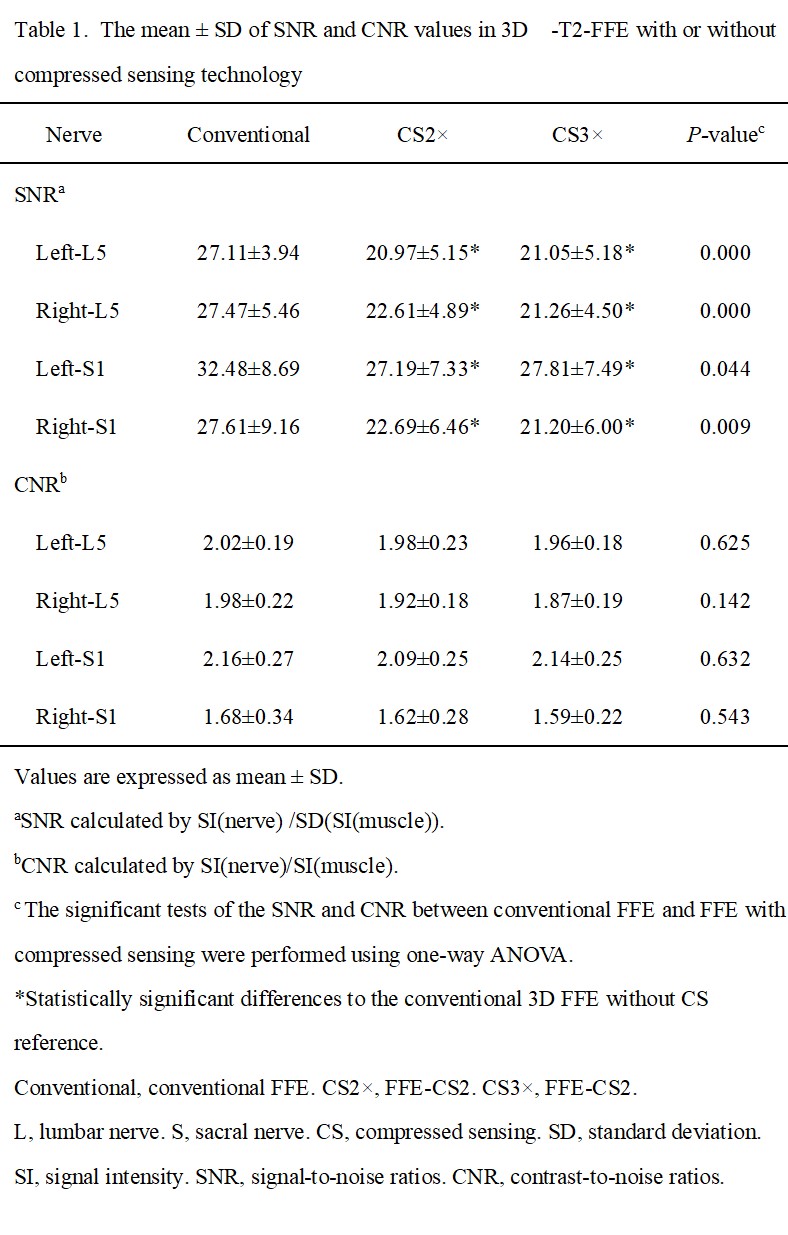
Compressed sensing technology can greatly
improve scanning speed at the small expense of a small SNR with unchanged CNR, morphological
measurements and subjective images evaluation score on lumbosacral plexus MR
imaging.
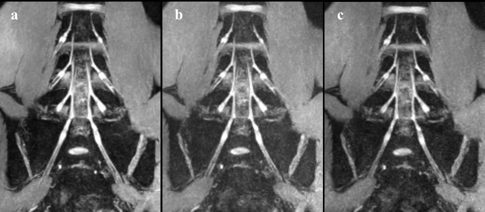
Figure 3. Comparison of the conventional FFE with the FFE-CS. LSP magnetic resonance
neurography images of a 26-year-old woman without lower back pain. The coronal conventional 3D FFE (a) serves as a reference
for the FFE-CS with factor CS2(b) and CS3(c). No difference can be observed among
the three types of FFE, all sequences showing compromised image quality. A
general noise enhancement could be observed in the FFE sequences with
compressed sense. The subjective image quality score for LSP given by the two radiologists was both 3 in the conventional and
compressed sensing sequences.