Ryckie George Wade1, Alexander Whittam2, Irvin Teh1, Gustav Andersson3, Fang-Cheng Yeh 4, Mikael Wiberg 3, and Grainne Bourke 1
1University of Leeds, Leeds, United Kingdom, 2Sheffield Teaching Hospitals, Sheffield, United Kingdom, 3Umeå University, Umeå, Sweden, 4University of Pittsburgh, Pittsburgh, PA, United States
1University of Leeds, Leeds, United Kingdom, 2Sheffield Teaching Hospitals, Sheffield, United Kingdom, 3Umeå University, Umeå, Sweden, 4University of Pittsburgh, Pittsburgh, PA, United States
In this systematic review and meta-analysis of the normal diffusion tensor imaging values for spinal roots of the brachial plexus, we show that the pooled mean FA of the roots was 0.36 (95% CI 0.34, 0.38) and the pooled mean MD of the roots was 1.51 x10⁻³ mm2/s (95% CI 1.45, 1.56).
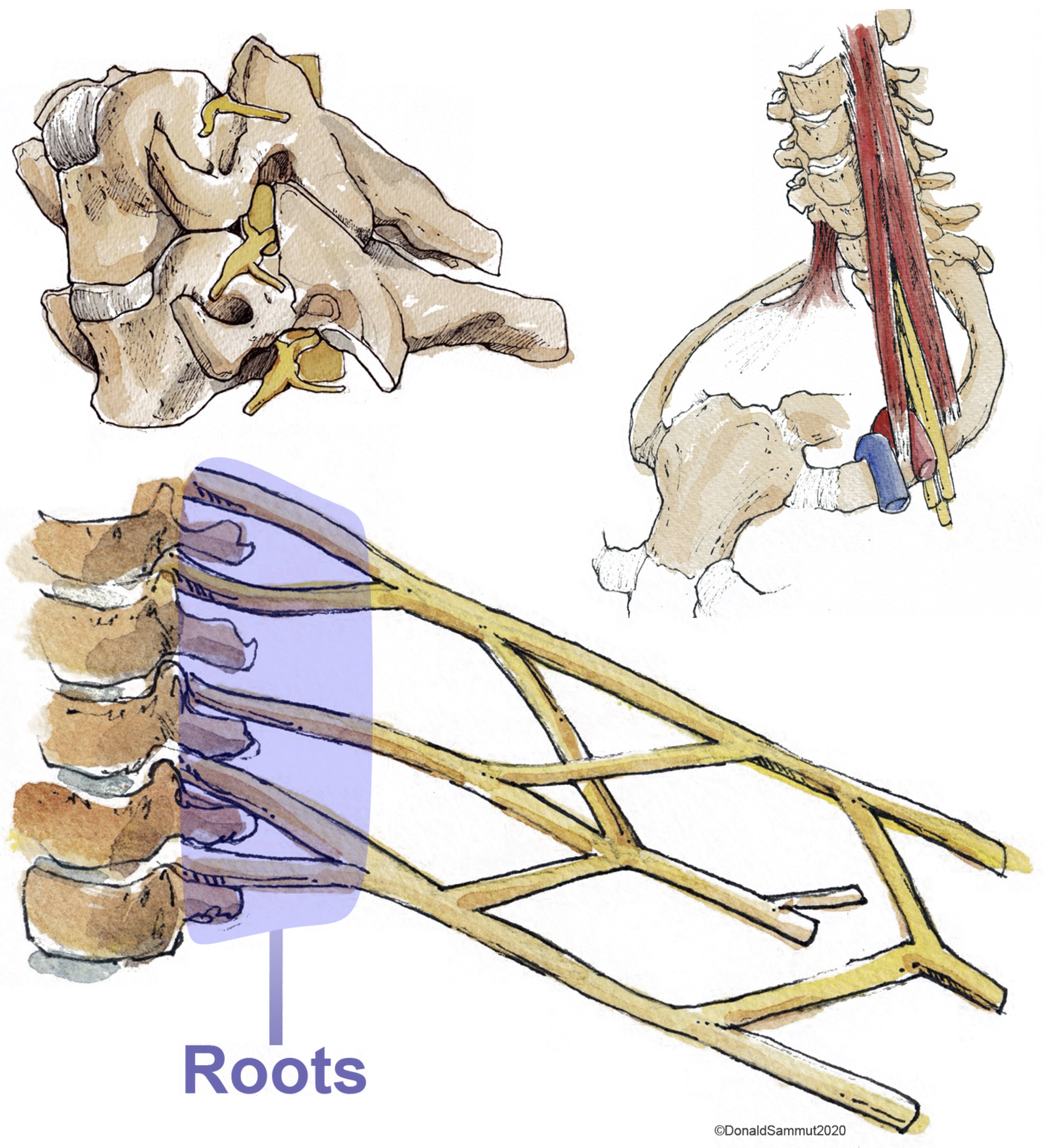
Figure 1. The roots of the brachial plexus emerging from the intervertebral foramina (upper left image) and their relationship to the scalene muscles and vasculature of the upper limb (upper right image). The lower image is a simplified schematic of the brachial plexus highlighting (in purple) the spinal roots. Reproduced with permission from Mr Donald Sammut.
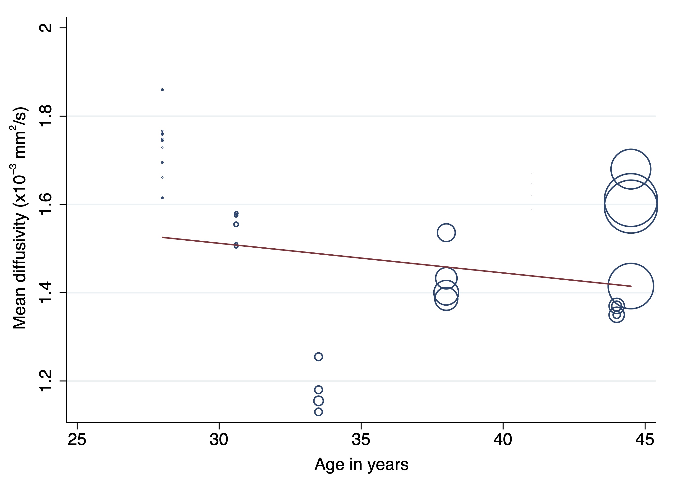
Figure 3. A scatterplot showing the negative association between the mean diffusivity of the roots of the brachial plexus and the mean age of adults in the included studies.