Stefano Tambalo1,2, Riccardo Pederzolli2, Andrea Spagnolo2, Lisa Novello1, and Jorge Jovicich1
1CIMeC, University of Trento, Trento, Italy, 2Department of Radiology, G.B. Rossi Hospital, University of Verona, Verona, Italy
1CIMeC, University of Trento, Trento, Italy, 2Department of Radiology, G.B. Rossi Hospital, University of Verona, Verona, Italy
We propose a tool that automatically detects “poor fat-suppression like” artifacts in diffusion MRI data. The tool indicates the specific image slices and volumes where the artifact is present, with high specificity (0.977) and sensitivity (0.889).
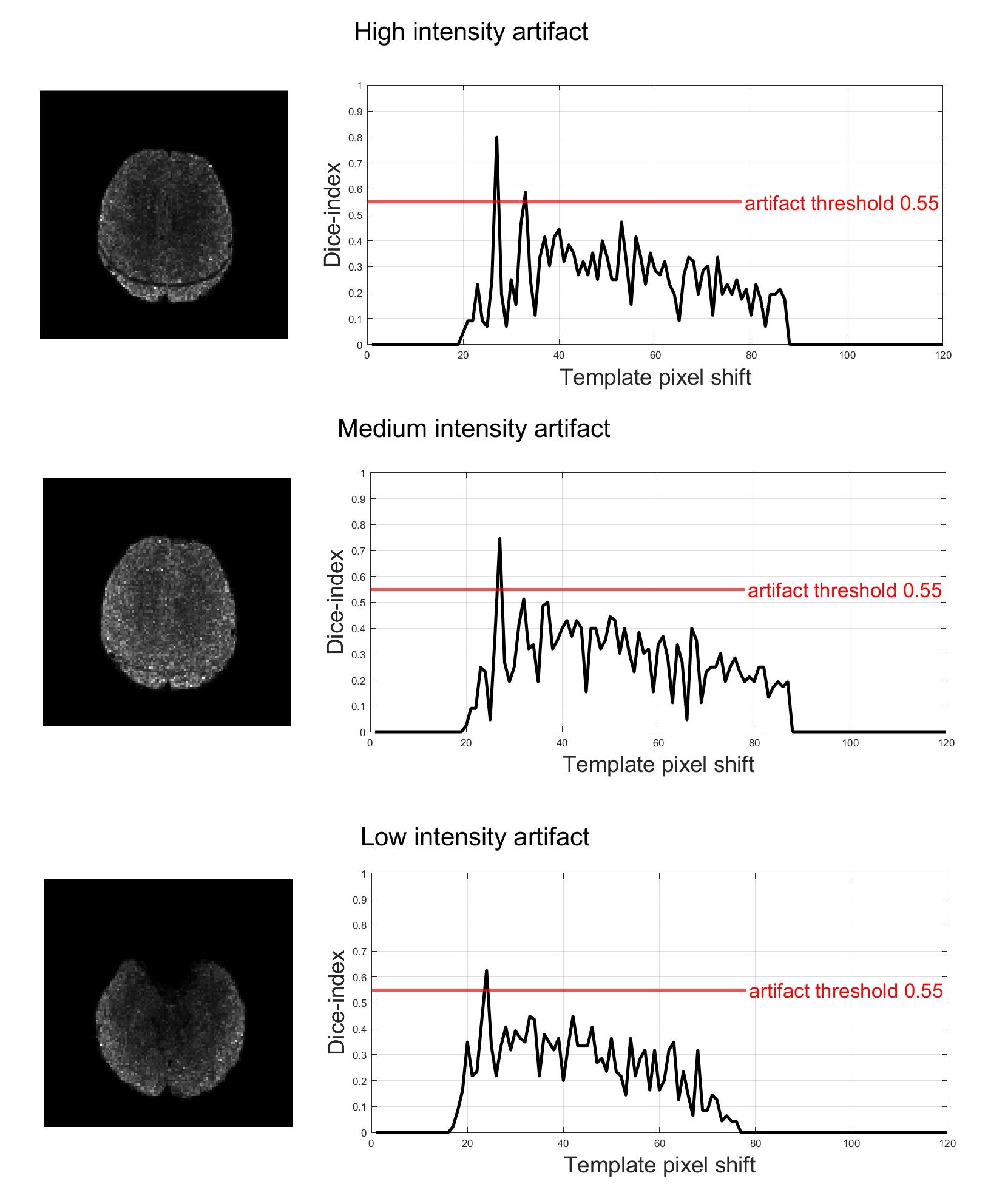
Figure 3. Representative images with different level of artifact intensity and corresponding Dice Index values as a function of posterior-anterior template shift. From top to bottom: high, medium, and low level. The prominent peak in each graph corresponds to the best-matching offset of template and artifact.
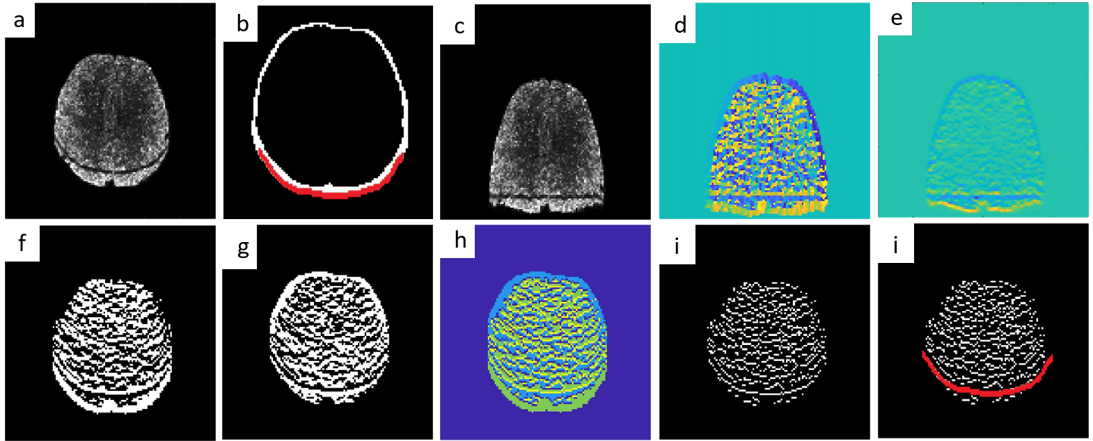
Figure 2. A brief overview of the tool processing steps from input image to artifact detection. a) input image; b) skull mask and template; straightened image; d,e) image gradient; f, g, h) image gradient merged and shifted for edge enhancement; i) base image for template matching; j) overlay of base image and template. A detailed description of each step is given in the text.