Thea Bautista1, Jonathan O'Muircheartaigh2,3,4,5, Joseph V Hajnal1,3, and J-Donald Tournier1,3
1Department of Biomedical Engineering, School of Biomedical Engineering and Imaging Sciences, King's College London, London, United Kingdom, 2Forensic & Neurodevelopmental Sciences, King's College London, London, United Kingdom, 3Centre for the Developing Brain, School of Biomedical Engineering and Imaging Sciences, King's College London, London, United Kingdom, 4Department of Perinatal Imaging & Health, School of Biomedical Engineering and Imaging Sciences, King's College London, London, United Kingdom, 5MRC Centre for Neurodevelopmental Dirorders, King's College London, London, United Kingdom
1Department of Biomedical Engineering, School of Biomedical Engineering and Imaging Sciences, King's College London, London, United Kingdom, 2Forensic & Neurodevelopmental Sciences, King's College London, London, United Kingdom, 3Centre for the Developing Brain, School of Biomedical Engineering and Imaging Sciences, King's College London, London, United Kingdom, 4Department of Perinatal Imaging & Health, School of Biomedical Engineering and Imaging Sciences, King's College London, London, United Kingdom, 5MRC Centre for Neurodevelopmental Dirorders, King's College London, London, United Kingdom
The subvoxel shifts method for the removal of Gibbs ringing artefacts can trivially be extended to 3D with simple modifications, and performs well on numerical phantom and in vivo data.
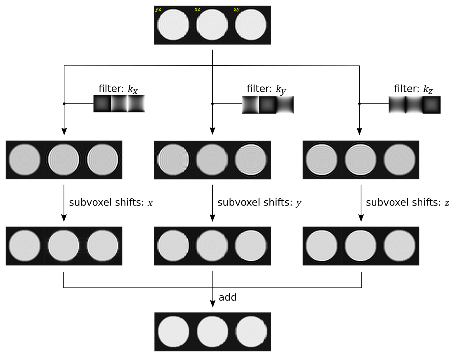
overview of the 3D subvoxel shifts approach. Each panel shows three orthogonal cuts through the 3D image. The approach is identical to the original 2D version, but applies the filtering and subvoxels shifts along all three spatial axes, with modified filters appropriate for the 3D case. For each axis, the image is Fourier filtered to attenuate high frequencies in the directions orthogonal to the axis of interest. Gibbs ringing removal is then applied using 1D subvoxel shifts along that axis. The resulting three images are then summed together to give the final output.
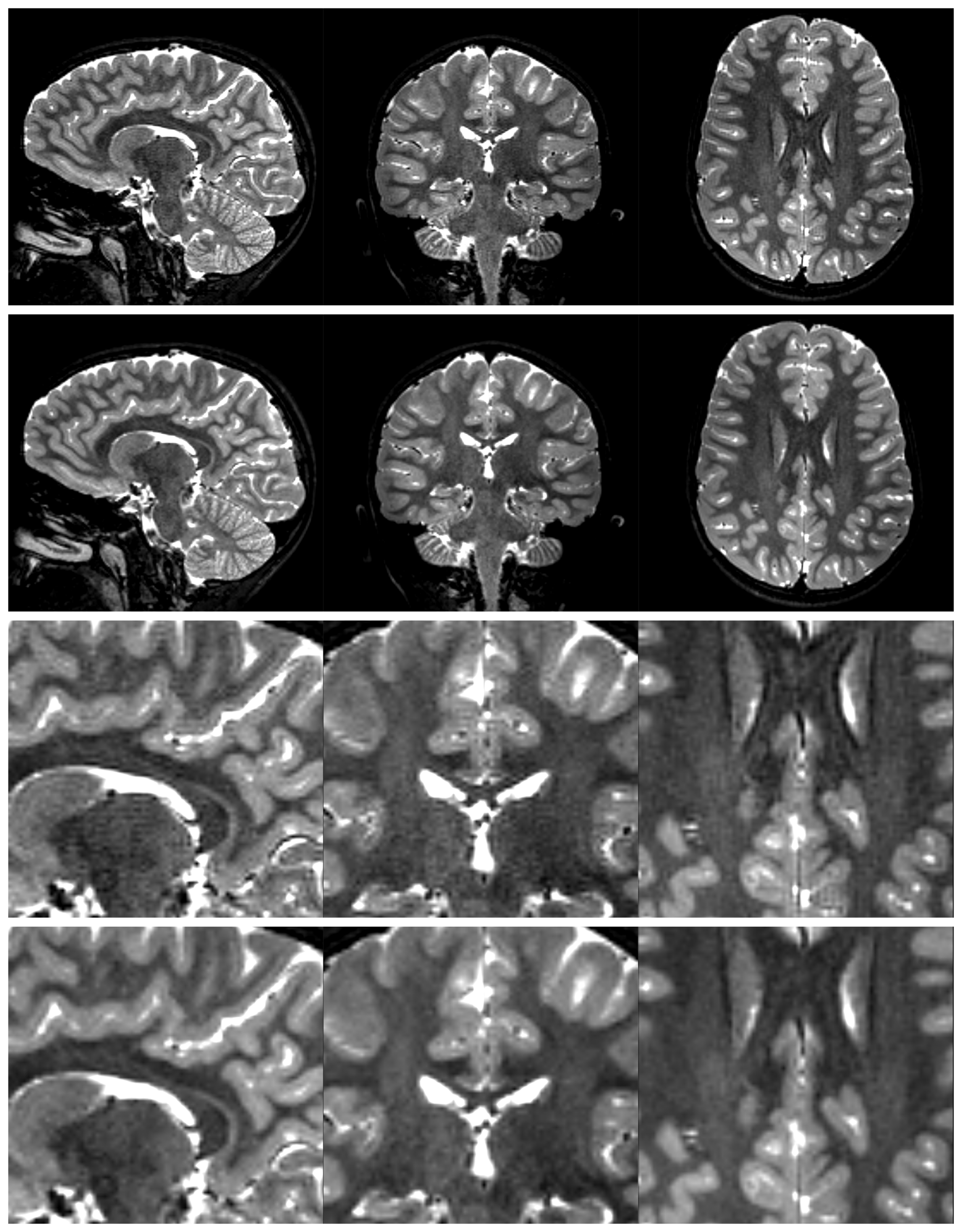
demonstration of the proposed method in practice, using a T2-weighted 3D acquisition (details). The top two rows show 3 orthogonal cuts through the original image with evident Gibbs ringing artefacts and the output of the proposed method. The bottom two rows show magnified sections of the same images for closer inspection.